PSEB 11th Class Biology Solutions Chapter 10 Cell Cycle and Cell Division

Punjab State Board PSEB 11th Class Biology Book Solutions Chapter 10 Cell Cycle and Cell Division Textbook Exercise Questions and Answers.

PSEB Solutions for Class 11 Biology Chapter 10 Cell Cycle and Cell Division

PSEB 11th Class Biology Guide Cell Cycle and Cell Division Textbook Questions and Answers

Question 1.
What is the average cell cycle span for a mammalian cell ?
Answer:
The average cell cycle span for a mammalian cell is 24 hours.

Question 2.
Distinguish cytokinesis from karyokinesis?
Answer:
Cytokinesis is the division of cytoplasm, whereas karyokinesis is the division of nucleus of the cell.

PSEB 11th Class Biology Solutions Chapter 10 Cell Cycle and Cell Division

Question 3.
Describe the events taking place during interphase.
Answer:
The interphase, though called the resting phase, is the time during which the cell is preparing for division by undergoing cell growth and DNA replication. The interphase is divided into three further phases
(i) G1 – phase (Gap 1)
(ii) S – phase (Synthesis)
(iii) G2 – phase (Gap 2)
G1 – phase corresponds to the interval between mitosis and initiation of DNA replication. During Ga-phase the cell is metabolically active and continuously grows but does not replicate its DNA. S or synthesis phase marks the period during, which, DNA synthesis or replication takes place. During this time, the amount of DNA per cell doubles.

During the G2 – phase, proteins are synthesised in preparation for mitosis, while cell growth continues.
Cells in this stage remain metabolically active but no longer proliferate unless called on to do so depending on the requirement of the organism.

Question 4.
What is G0 (quiescent phase) of cell cycle?
Answer:
G0 is the quiescent stage of the cell cycle. It is also known as inactive stage of the cell cycle. Cells in this stage remain metabolically remain active, but no longer proliferate unless called on to do so depending on the requirement of the organisms.

Question 5.
Why is mitosis called equational division?
Answer:
Mitosis is the kind of cell division in which daughter cells have the same number and kind of chromosomes as that of parent cell. Mitosis is therefore, also known as equational division.

Question 6.
Name the stage of cell cycle at which one of the following events occur:
(i) Chromosomes are moved to spindle equator.
(ii) Centromere splits and chromatids separate.
(iii) Pairing between homologous chromosomes takes place.
(iv) Crossing over between homologous chromosomes takes place.
Answer:
(i) Prophase,
(ii) Anaphase,
(iii) Zygotene stage of meiosis-I,
(iv) Pachytene stage.

PSEB 11th Class Biology Solutions Chapter 10 Cell Cycle and Cell Division

Question 7.
Describe the following:
(i) Synapsis
(ii) Bivalent
(iii) Chiasmata
Draw a diagram to illustrate your answer.
Answer:
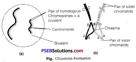
(i) Synapsis: During meiosis-I, the process of pairing of two homologous chromosomes is known as synapsis. It is so exact that pairing is not merely* between corresponding chromosomes but between corresponding individual units.

(ii) Bivalent: The complex formed by a pair of synapsed homologous chromosomes is called a bivalent or a tetrad. It consists of four chromatids.

(iii) Chiasmata: The chiasmata formation is the indication of completion of crossing over and beginning of separation of chromosomes. The chiasma is formed when the chromosomal parts begin to repel each other except in the region where these are in contact. Thus, chiasmata formation is necessary for the separation of homologous chromosomes.
PSEB 11th Class Biology Solutions Chapter 10 Cell Cycle and Cell Division 1

Question 8.
How does cytokinesis in plant cells differ from that in animal cells?
Answer:
Cytokinesis in an Animal Cell: It occurs by a process called cleavage. A cleavage furrow appears and at the site of the cleavage furrow the cytoplasm has a ring of microfilaments made of actin associated with molecules of the protein myosin. This ring of proteins will contract causing the furrow to deepen, which will then in turn pinch the cell into two separate cells.

Cytokinesis in a Plant Cell: In a plant cell, vesicles containing cell wall material collect at the middle of the parent cell. They will fuse and form a membranous cell plate. This plate will grow outward and it accumulates more cell wall material. Eventually, the plate will fuse with the plasma membrane and the cell plate contents will join the parental cell wall. This results in two different daughter cells.

Question 9.
Find examples where the four daughter cells from meiosis are equal in size and where they are found unequal in size.
Answer:
The four daughter haploid cells may or may not be equal in size at the end of meiosis-II with telophase-II

PSEB 11th Class Biology Solutions Chapter 10 Cell Cycle and Cell Division

Question 10.
Distinguish anaphase of mitosis from anaphase-1 of meiosis. Ans. Differences between anaphase of mitosis and anaphase-I of meiosis.
Answer:

Anaphase of Mitosis Anaphase-I of Meiosis
1. Each chromosome arranged at the metaphase plate is split simultaneously and the two daughter chromatids, begin to migrate towards the two opposite poles. The spindle fibres contract and pull the centromeres of homologous chromosomes towards the opposite poles.
2. The centromere of each chromosome is towards the pole with arms of chromosome trailing behind. The centromere is not divided, so half of the chromosomes of parent nucleus go to one pole and the remaining half in the opposite pole.
3. In this stage

(a) centromeres split and chromatids separate.

(b) chromatids move to opposite poles.

In this stage

(a) homologous chromosomes separate.

(b) sister chromatids remain associated at their centromere.

Question 11.
List the main differences between mitosis and meiosis.
Answer:

Mitosis Meiosis
1. The division occurs in somatic cells. It occurs in reproductive cells.
2. It is a single division. It is a double division.
3. The daughter cells resemble each other as well as their mother cell. The daughter cells neither resemble one another nor their mother cell.
4. Replication of chromosomes occurs before every mitotic division. Replication of chromosomes occurs only once though meiosis is a double division.
5. Mitosis does not introduce variations. Meiosis introduces variations.
6. Mitosis is required for growth, repair and healing. Meiosis is involved in sexual reproduction.

Question 12.
What is the significance of meiosis?
Answer:
Meiosis is the mechanism by which conservation of specific chromosome number of each species is achieved across generations in sexually reproducing organisms.
It also increases the genetic variability in the population of organisms from one generation to the next. Variations are very important for the process of evolution.

PSEB 11th Class Biology Solutions Chapter 10 Cell Cycle and Cell Division

Question 13.
Discuss with your teacher about:
(i) haploid insects and lower plants where cell-division occurs, and
(ii) some haploid cells in higher plants where cell-division does
not occur.
Answer:
(i) In some insects and lower plants fertilization is immediately followed by zygotic meiosis, which leads to the production of haploid organisms. This type of life cycle is known as haplontic life cycle.

(ii) The phenomenon of polyploidy can be observed in some haploid cells in higher plants in which cell division does not occur. Polyploidy is a state in which cells contain multiple pairs of chromosomes than the basic set. Polyploidy can be artificially induced in plants by applying colichine to cell culture.

Question 14.
Can there be mitosis without DNA replication in S-phase?
Answer:
No, DNA replication is required for doubling the chromosome number.

Question 15.
Can there be DNA replication without cell division?
Answer:
Yes, DNA replication takes place during the synthesis stage of interphase in the cell cycle. It is totally independent of cell division. After G2 -phase a cell may or may not enter into the M-phase.

PSEB 11th Class Biology Solutions Chapter 10 Cell Cycle and Cell Division

Question 16.
Analyse the events during every stage of cell cycle and notice how the following two parameters change?
(i) Number of chromosomes (n) per cell
(ii) Amount of DNA content (C) per cell.
Answer:
(i) The number of chromosomes (n) is double in the prophase, metaphase and anaphase stage of an organism. In the telophase stage, during daughter cell formation, the number of chromosomes get reduced to half.

(ii) Amount of DNA content (C) per cell also changes during different phases of cell division. It is double in the prophase, metaphase and anaphase stage of an organism. In the telophase stage, during daughter cell formation, the number of chromosomes get reduced to half than that of their parents. ’
PSEB 11th Class Biology Solutions Chapter 14 Respiration in Plants

Punjab State Board PSEB 11th Class Biology Book Solutions Chapter 14 Respiration in Plants Textbook Exercise Questions and Answers.

PSEB Solutions for Class 11 Biology Chapter 14 Respiration in Plants

PSEB 11th Class Biology Guide Respiration in Plants Textbook Questions and Answers

Question 1.
Differentiate between:
(a) Respiration and Combustion
(b) Glycolysis and Krebs’ cycle
(c) Aerobic respiration and Fermentation
Answer:
(a) Differences between Respiration and Combustion

Respiration Combustion
1. It is the breakdown of complex compounds through oxidation within the cells, leading to release of considerable amount of energy. Combustion is the complete burning of glucose, which produces CO2 and H2O and yields energy which is given out as heat.
2. It is a controlled biochemical process. It is an uncontrolled physicochemical process.
3. Many chemical bonds break simultaneously by releasing large amounts of energy. Chemical bonds break one after another to release energy.
4. Enzymes are involved. Enzymes are not involved.

(b) Differences between Glycolysis and Krebs’ Cycle

Glycolysis Krebs’ Cycle
1. It is also called as Embden Meyerhof-Parnas (EMP) pathway. It is also known as citric acid cycle or tricarboxylic acid cycle.
2. It occurs in the cytosol of prokaryotes as well as eukaryotes. In eukaryotes, it occurs in matrix of mitochondria, and in prokaryotes, it occurs in cytoplasm.
3. It starts with the oxidation of glucose. It starts with the oxidation of pyruvic acid.
4. It is an enzyme-controlled 10 steps process by which glucose, fructose or sucrose is reduced to form pyruvic acid with the production of ATP and NADH2. It involves 8 steps to oxidize two molecules of acetyl CO-A

(c) Differences between Aerobic Respiration and Fermentation

Aerobic Respiration Fermentation
1. The presence of O2 is required for complete oxidation of organic substances. Incomplete oxidation of glucose occurs in the absence of oxygen.
2. It releases CO2, water, and a large amount of energy present in the substrate. Pyruvic acid is converted to CO2 and ethanol and less amount of energy is released.
3. Only two molecules of ATP are produced. Many molecules of ATP are produced.
4. NADH is oxidized to NAD+ slowly. This reaction is very vigorous under aerobic respiration.

PSEB 11th Class Biology Solutions Chapter 14 Respiration in Plants

Question 2.
What are respiratory substrates? Name the most common respiratory substrate.
Answer:
The compounds oxidized during the process of respiration are called respiratory substrates. Carbohydrates, especially glucose, act as respiratory substrates. Fats, proteins, and organic acids also act as respiratory substrates.

Question 3.
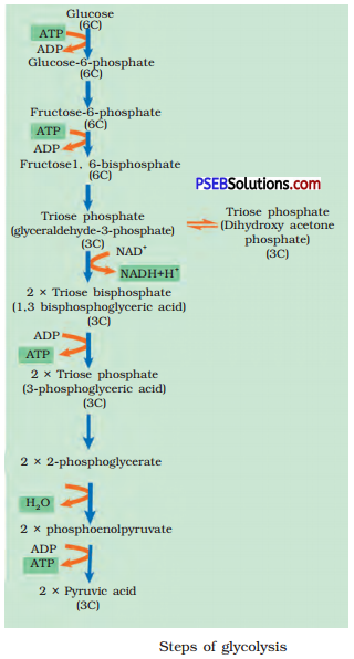
Give the schematic representation of glycolysis.
Answer:
Glycolysis

  • The scheme of reactions was given by Embden, Meycrhof and Pumas, hence it is also called EMP Pathway; it occurs in the cytoplasm of cells.
  • In this process, glucose is partially oxidized/ converted into two molecules of pyruvic acid.
  • Glucose is phosphorylated with the help of ATP into Glucose-6-Phosphate, catalyzed by the enzyme hexokinase.
  • Glucose-6-Phosphate is converted into its isomer, Fructose-6-Phosphate, catalyzed by isomerase.
  • Fructose-6-Phosphate is then phosphorylated with the use of ATP into Fructose 1,6-bisphosphate, catalyzed by
    phosphofructokinase.
  • Fructose 1,6-bisphosphate is split into one molecule of 3-pho sphoglyceraldchyd and one molecule of
    dihydroxy-acetone phosphate; these two products are interconvertible.
  • 3-phosphoglyceraldehyde is oxidized to 1, 3 bi phosphoglycerate, where NAD is reduced to NADH.
  • 1,3-bisphosphoglycerate is split into 3-phosphoglycerate along with the formation of ATP, catalyzed by phosphoglycerate kinase.

PSEB 11th Class Biology Solutions Chapter 14 Respiration in Plants 1

  • 3-Phosphoglycerate is subsequently converted into 2-phosphoglycerate and then into Phosphoenol Pyruvate (PEP).
  • PEP is converted into pyruvate along with the formation of ATP, catalyzed by the enzyme pyruvate kinase.

In this pathway, ATP molecules are formed in two ways :

  1. Direct/substrate phosphorylation of ADP to ATP.
  2. Oxidation of NADH to NAD ’ and ATP formation through electron transport (oxidative phosphorylation).

Four molecules of ATP are formed directly in the following steps:

  • When 1, 3 bi phosphoglyceric acids is converted into 3-phosphoglyceric acid and
  • When phosphoenolpyruvate is converted into pyruvic acid.

Two molecules of ATP are consumed, one in each of the following steps:

  1. Phosphorylation of glucose to glucose-6- phosphate and
  2. Conversion of fructose-6-phosphate into fructose 1, 6 bisphosphates.

When 3-phosphoglyceraldehyde (PGAL) is converted into 1, 3 bi phosphoglycerates, two redox equivalents are removed in the form of two hydrogen atoms from PGAL and transferred to NAD+, which is reduced to NADH + H+.
Oxidation of two molecules of NADH to NAD yields six molecules of ATP during electron transport. The end products of glycolysis are two molecules each of pyruvic acid, NADH, and ATP.

PSEB 11th Class Biology Solutions Chapter 14 Respiration in Plants

Question 4.
What are the main steps in aerobic respiration? Where does it take place?
Answer:
The main steps in aerobic respiration are as follows :

  • Glycolytic breakdown of glucose into pyruvic acid.
  • Oxidative decarboxylation of pyruvic acid to acetyl Co-A (acetyl coenzyme-A).
  • Krebs’ cycle.
  • Terminal oxidation and phosphorylation in respiratory chain.
    It occurs inside the mitochondrial matrix.

Question 5.
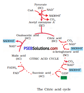
Give the schematic representation of an overall view of Kreb’s cycle.
Answer:
Tricarboxylic Acid Cycle or Kreb’s Cycle:

  • It is known as tricarboxylic acid cycle, since the first product, citric acid (hence citric acid cycle) is a tricarboxylic acid (has three COOH groups).
  • The sequence of reactions in citric acid cycle was first followed by Sir Hans Kreb; hence it is also known as Kreb’s cycle.
  • During this cycle of reactions, 3 molecules of NAD and one molecule of FAD are reduced to NADH and FADH respectively.
  • Respiration in Plants 115:
  • One ATP molecule is produced directly from GTP formed by substrate-level phosphorylation.

The reactions of aerobic oxidation of Pyruvic acid can be summed up as :
Pyruvic acid + 4NAD+ + FAD+ + 2H2O + ADP + Pi → 3CO2 + 4NADH + 4H+ + FADH2 + ATP
PSEB 11th Class Biology Solutions Chapter 14 Respiration in Plants 2

Question 6.
Explain ETS.
Answer:
Electron Transport System (ETS):

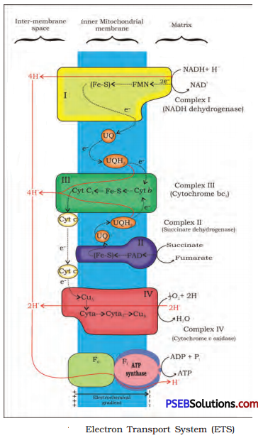
  • ETS occurs in the electron transport particles (ETP) on the inner surface of the inner membrane of mitochondria.
  • NADH formed in glycolysis and citric acid cycle are oxidized by NADH-dehydrogenase (complex I) and the electrons are transferred to ubiquinone, via FMN.
  • Ubiquinone also receives reducing equivalents via FADH generated during the oxidation of succinate by succinate dehydrogenase (complex II).
  • The reduced ubiquinone called ubiquinol is then oxidized by transfer of electrons to cytochrome c, via cytochrome be bc1 -complex (complex III).
  • Cytochrome c acts as a mobile carrier between complex III and complex IV.
  • Complex IV refers to cytochrome c oxidase complex containing cytochromes a and a3 and two copper centres.
  • When the electrons are shunted over the carriers via complex I to IV in the electron transport chain, they are coupled to ATP synthetase (complex V) for the formation of ATP from ADP and IP.
  • Oxygen functions as the terminal acceptor of electrons and is reduced to water along with the hydrogen atoms.

PSEB 11th Class Biology Solutions Chapter 14 Respiration in Plants 3

Question 7.
Distinguish between the following:
(a) Aerobic respiration and anaerobic respiration.
(b) Glycolysis and fermentation.
(c) Glycolysis and citric acid cycle.
Answer:
(a) Differences between Aerobic and Anaerobic Respiration

Aerobic Respiration Anaerobic Respiration
1. Presence of oxygen is essential. Occurs without oxygen.
2. Complete oxidation of substrates occurs into CO2 and H2O. Incomplete degradations of substrates.
3. End products are non-toxic. End products are toxic when accumulated in large amount.
4. Involves glycolysis, Krebs’ cycle, and terminal oxidation. Involves only glycolysis followed by incomplete breakdown of pyruvic acid.
5. 36 ATP molecules are produced from each glucose molecule. Only two molecules of ATP are produced from each glucose molecule.

(b) Differences between Glycolysis and Fermentation:

Glycolysis Fermentation
1. it occurs in cytoplasm of the cell and in all living things. it occurs in yeast and muscle cells in animals when oxygen is not sufficient for cellular respiration.
2. Glucose undergoes partial oxidation to form two molecules of pyruvic acid. The enzyme-like pyruvic acid decarboxylase and alcohol dehydrogenase catalyze these reactions.
3. The end products are CO2, H2O and energy. The end products are CO2 and ethanol and in animal cells lactic acid is released.

(c) Differences between Glycolysis and Krebs’ Cycle

Glycolysis Krebs’ Cycle
1. It is also called as Embden Meyerhof-Parnas (EMP) pathway. It is also known as citric acid cycle or tricarboxylic acid cycle.
2. It occurs in the cytosol of prokaryotes as well as eukaryotes. In eukaryotes, it occurs in matrix of mitochondria and in prokaryotes, it occurs in cytoplasm.
3. It starts with the oxidation of glucose. It starts with the oxidation of pyruvic acid.
4. It is an enzyme-controlled 10 steps process by which glucose, fructose or sucrose is reduced to form pyruvic acid with the production of ATP and NADH2. It involves 8 steps to oxidize two molecules of acetyl CO-A

Question 8.
What are the assumptions made during the calculation of net gain of ATP?
Answer:
The calculations of net gain of ATP can be made only on certain assumptions :

  • There is a sequential, orderly pathway functioning, with one substrate forming the next and with glycolysis, TCA cycle and ETS pathway following one after another.
  • The NADH synthesized in glycolysis is transferred into the mitochondria and undergoes oxidative phosphorylation.
  • None of the intermediates in the pathway are utilised to synthesize any other compound.
  • Only glucose is being respired-no other alternative substrates are entering in the pathway at any of the intermediary stages.

But this kind of assumptions are not really valid in a living system. All pathways work simultaneously and do not take place one after another. Substrates enter the pathways and are withdrawn from it as end when necessary; ATP is utilized as and when needed; enzymatic rates are controlled by multiple means. In overall steps, there is a net gain of 36 ATP molecules during aerobic respiration of one molecule of glucose.

Question 9.
Discuss ‘the respiratory pathway is an amphibolic pathway.’
Answer:
Aniphibolic Pathway: Glucose is the favored substrate for respiration. All carbohydrates are usually first converted into glucose before they are used for respiration. Other substrates can also be respired but then they do not enter the respiratory pathway at the first step.

Since respiration involves breakdown as well as synthesis of substrates, the respiratory process involves both catabolism and anabolism. That is why respiratory pathway is considered to be an amphibolic pathway rather than as a catabolic one.

Question 10.
Define RQ. What is its value for fats?
Answer:
Respiratory quotient (RQ) or respiratory ratio can be defined as the ratio of the volume of CO2 evolved to the volume of O2 consumed during respiration. The value of respiratory quotient depeñds on the type of respiratory substrate. Its value is one for carbohydrates. However, it is always less than one for fats as fats consume more oxygen for respiration than carbohydrates.

It can be illustrated through the example of tripalmitin fatty acid, which consumes 145 molecules of O2 for respiration while 102 molecules of CO2 are evolved.The RQ value for tripalmitin is 0.7.

Question 11.
What is oxidative phosphorylation?
Answer:
It refers to the formation of ATP in the mitochondria, utilizing the energy obtained by the oxidation of organic molecules.

Question 12.
What is the significance of step-wise release of energy in respiration?
Answer:
During oxidation within a cell, all the energy contained in respiratory substrates is not released free in a single step. It is released in a series of slow step-wise reactions controlled by enzymes and it is trapped as chemical energy in the form of ATP. The significance of this step-wise release of energy is that some energy is used to synthesize ATP. This ATP is stored for the later utilization wherever required. Hence, ATP acts as the energy currency of cell. The energy

stored in ATP can be utilized for following:

  • In various energy-requiring processes of organisms.
  • The carbon skeleton produced during respiration is used as precursors for the synthesis of other molecules.

PSEB 11th Class Biology Solutions Chapter 9 Biomolecules

Punjab State Board PSEB 11th Class Biology Book Solutions Chapter 9 Biomolecules Textbook Exercise Questions and Answers.

PSEB Solutions for Class 11 Biology Chapter 9 Biomolecules

PSEB 11th Class Biology Guide Biomolecules Textbook Questions and Answers

Question 1.
What are macromolecules? Give examples.
Answer:
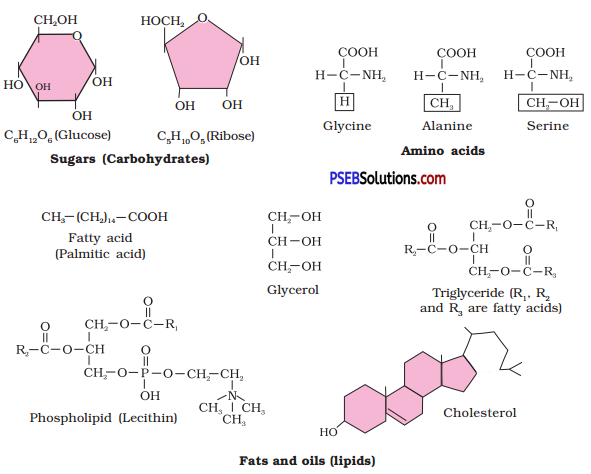
Chemical compounds, which are found in the acid insoluble fraction are called macromolecules or biomacromolecules. For example, proteins, lipids and carbohydrate, etc.

Question 2.
Illustrate a glycosidic, peptide and a phosphodiester bond.
Answer:
Glycosidic Bond: A glycosidic bond is a type of functional group that joins a carbohydrate (sugar) molecule to another group, which may or may not be another carbohydrate.
PSEB 11th Class Biology Solutions Chapter 9 Biomolecules 1
Peptide Bond: A peptide bond (amide bond) is a chemical bond formed between two molecules when the carboxyl group of one molecule reacts with the amine group of the other molecule, thereby releasing a molecule of water (H20).
H2O.
PSEB 11th Class Biology Solutions Chapter 9 Biomolecules 2
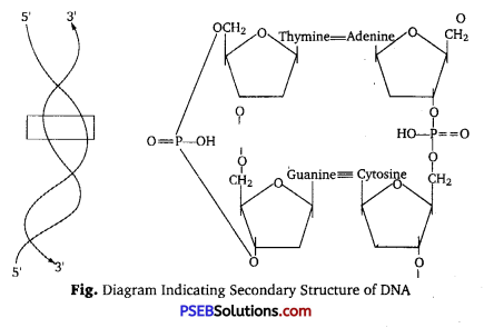
Phosphodiester Bond: A phosphodiester bond is a group of strong covalent bonds between a phosphate group and two other molecules over two ester bonds. In DNA and RNA, the phosphodiester bond is the linkage between the 3′ carbon atom of one sugar molecule and the 5’carbon of another, deoxyribose in DNA and ribose in RNA.
PSEB 11th Class Biology Solutions Chapter 9 Biomolecules 3
PSEB 11th Class Biology Solutions Chapter 9 Biomolecules

Question 3.
What is meant by tertiary structure of proteins?
Answer:
Tertiary Structure of Protein; The overall shape of a single protein molecule; the spatial relationship of the secondary structures to one another. Tertiary structure is generally stabilized by non-local interactions, most commonly the formation of a hydrophobic core, but also through salt bridges, hydrogen bonds, disulfide bonds,1 and even post-translational modifications. The term “tertiary structure” is often used as synonymous with the term fold. The tertiary structure is what controls the basic function of the protein.

Question 4.
Find and write down structures of 10 interesting small molecular weight biomolecules. Find if there is any industry which manufactures the compounds by isolation. Find out who are the buyers?
Answer:
PSEB 11th Class Biology Solutions Chapter 9 Biomolecules 4
PSEB 11th Class Biology Solutions Chapter 9 Biomolecules 5

Question 5.
Proteins have primary structure. If you are given a method to know which amino acid is at either of the two termini (ends) of a protein, can you connect this information to purity or homogeneity of a protein?
Answer:
The sequence of amino acids, i.e., the positional information in a protein which is the first amino acid, which is second and so on is called the primary structure of a protein. The first amino acid is also called as N-terminal amino acid. The last amino acid is called the C-terminal amino acid. Yes, we can connect this information to purity or homogeneity of a protein. Based on number of amino and carboxyl groups, there are acidic (e.g., glutamic acid), basic (lysine) and neutral (valine) amino acids, proteins may be acidic, basic and neutral.

PSEB 11th Class Biology Solutions Chapter 9 Biomolecules

Question 6.
Find out and make a list of proteins used as therapeutic agents. Find other applications of proteins (e. g, cosmetics etc.)
Answer:
Some proteins and their functions are as follows:

Proteins Functions
1. Collagen Intercellular ground substance
2. Trypsin Enzyme
3. Insulin Hormone
4. Antibody Fights against infections
5. Receptors Sensory reception (example-taste)
6. Glut-4 Enables glucose transport in cells
7. Keratolytic protein Used to soften hard skin
8. Egg protein Used for skin tightening

Question 7.
Explain the composition of triglyceride.
Answer:
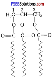
Triglycerides are composed of two types of molecules, i.e., glycerol i (3 carbon molecules) and fatty acids which attach to the glycerol at the alcohol unit. The following is a structural representation of a triglyceride at the molecular level.
PSEB 11th Class Biology Solutions Chapter 9 Biomolecules 6
Fatty acids are chains of hydrocarbons 4-22 (or more) carbons a long with a carboxyl group at one end. If each carbon has two hydrogen atoms, the fatty acid is saturated. If two carbon atoms are double-bonded, so that there is less hydrogen in the fatty acid, it is unsaturated (monounsaturated). If more than two carbon atoms are unsaturated, the fatty acid is polyunsaturated.

Question 8.
Can you describe what happens when milk is converted into curd or yoghurt, from your understanding of proteins.
Answer:
Milk contains a protein called casein. This protein gives milk its characteristic white colour. It is of high nutritional value because it contains all the essential amino acids required by man’s body. The curd forms because of the chemical reaction between lactic acid bacteria and casein. When curd is added to milk, the lactic acid bacteria present in it cause coagulation of casein and thus, convert it into curd.

Question 9.
Can you attempt building models of biomolecules using commercially available atomic models (Ball and Stick models).
Answer:
Yes, we can make models of biomolecules using commercially available atomic models.

PSEB 11th Class Biology Solutions Chapter 9 Biomolecules

Question 10.
Attempt titrating an amino acid against a weak base and discover the number of dissociating (ionisable) functional groups in the amino acid.
Answer:
When an amino acid is titrated against a weak base, it dissociates and gives two functional groups:
(i) -COOH group (carboxylic group)
(ii) Amino group (NH2/sub>)

Question 11.
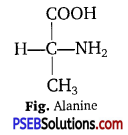
Draw the structure of the amino acid, alanine.
Answer:
PSEB 11th Class Biology Solutions Chapter 9 Biomolecules 7

Question 12.
What are gums made of? Is fevicol different?
Answer:
Gums are made of carbohydrates, i. e., L-rhamnose, D-galactose and D-galacturonic acid, etc. Fevicol is different from natural gums. It is a synthetic product.

Question 13.
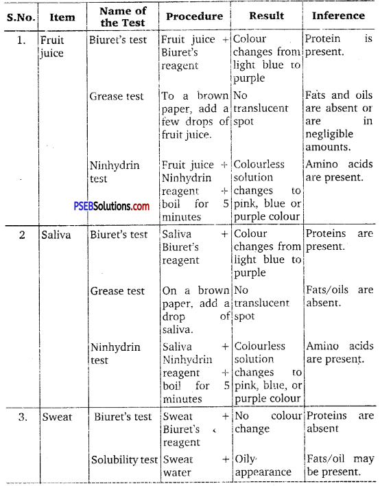
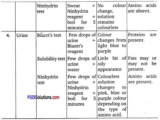
Find out a qualitative test for proteins, fats and oils, amino acids and test any Fruit juice, saliva, sweat and urine for them.
Answer:

  • Test for Proteins: Biuret test if Biuret’s reagent added to protein, then the colour of the reagent changes light blue to purple.
  • Test for Fats and Oils: Grease or test.
  • Test for Amino Acids: Ninhydrin test. If Ninhydrin reagent is added to the solution, then the colourless solution changes to pink, blue or purple depending upon the amino acid.

PSEB 11th Class Biology Solutions Chapter 9 Biomolecules 8
PSEB 11th Class Biology Solutions Chapter 9 Biomolecules 9

PSEB 11th Class Biology Solutions Chapter 9 Biomolecules

Question 14.
Find out how much cellulose is made by all the plants in the biosphere and compare it with how much of paper is manufactured by man and hence what is the consumption of plant material by man annually. What a loss of vegetation!
Answer:
Approximately, 100 billion tonnes of cellulose are made per year by all the plants in the biosphere and it takes 17 full grown trees to make one ton of paper. Trees are also used to fulfil the other requirements of man such as for timber, food, medicines, etc. Hence, it is difficult to calculate the annual consumption of plant material by man.

Question 15.
Describe the important properties of enzymes.
Answer:
Important properties of enzymes are given below :

  • Enzymes are proteins which catalyse biochemical reactions in the cells.
  • They are denatured at high temperatures.
  • Enzymes generally function in a narrow range of temperature and pH. Each enzyme shows its highest activity at a particular temperature and pH called the optimum temperature and optimum pH.
  • With the increase in substrate concentration, the velocity of the eyzymatic reaction rises at first. The reaction ultimately reaches a maximum velocity (Vmax) which is not exceeded by any further rise in concentration of the
    substrate.
  • The activity of an enzyme is also sensitive to the presence of specific chemicals that bind to the enzyme.
  • Enzymes are substrate specific in their action.

PSEB 11th Class Biology Solutions Chapter 15 Plant Growth and Development

Punjab State Board PSEB 11th Class Biology Book Solutions Chapter 15 Plant Growth and Development Textbook Exercise Questions and Answers.

PSEB Solutions for Class 11 Biology Chapter 15 Plant Growth and Development

PSEB 11th Class Biology Guide Plant Growth and Development Textbook Questions and Answers

Question 1.
Define growth, differentiation, development, dedifferentiation, redifferentiation, determinate growth, meristem and growth rate.
Answer:
Growth: It is an irreversible permanent increase in size of an organ or its parts or even of an individual cell.
Determinate Growth: Although growth in most of the plant parts is unlimited, certain parts grow up to a certain level and then stop growing. This kind of growth is known as determinate growth.

Development: It is the process of whole series of changes which an organism goes through during its life cycle.
Meristem: The cells of which the capacity to divide and self-perpetuate.

Growth Rate: The increased growth per unit time is termed as growth rate. Thus, rate of growth can be expressed mathematically. An organism or a part of the organism can produce more cells in a variety of ways. The growth rate shows an increase that may be arithmetic or geometrical.

Differentiation: The cells derived from root apical and shoot-apical meristems and cambium differentiate and mature to perform specific functions. This act leading to maturation is termed as differentiation.

Dedifferentiation: The living differentiated cells, that by now have lost the capacity to divide can regain the capacity of division under certain conditions. This phenomenon is termed as dedifferentiation. For example, formation of meristems; interfascicular cambium, and cork cambium from fully differentiated parenchyma cells. Redifferentiation: While undergoing dedifferentiation plant cells once again lose their capacity to divide and become mature. This process is called redifferentiation.

Question 2.
Why is not anyone parameter good enough to demonstrate growth throughout the life of a flowering plant?
Answer:
Anyone parameter is not good enough to demonstrate growth throughout the life of a flowering plant because the plants exhibit different types of growth during different stages of their life cycle. In the seedling stage, they are in state of active cell division (i.e., mitotic divisions), then they undergo active cell enlargement stage during growing stage. In the reproductive or flowering stage of their life cycle, they exhibit reductional divisions. Finally, after the formation of various organs, they undergo cell differentiation or get matured.

PSEB 11th Class Biology Solutions Chapter 15 Plant Growth and Development

Question 3.
Describe briefly:
(a) Arithmetic growth
(b) Geometric growth
(c) Sigmoid growth curve
(d) Absolute and relative growth rates
Answer:
(a) Arithmetic Growth: In arithmetic growth, following mitotic cell division only One daughter cell continues to divide while the other differentiates and matures. Example is a root elongating at a constant rate.

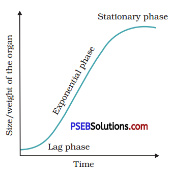
(b) Geometric Growth: In geometrical growth, in most systems, the initial growth is Size of slow (lag phase), and it organ increases rapidly thereafter at an exponential rate (log or exponential phase). Here, both the progeny cells following mitotic cell division retain the ability to divide and continue to do so.
PSEB 11th Class Biology Solutions Chapter 15 Plant Growth and Development 1
(c) Sigmoid Growth Curve: If we plot the parameter of growth against time, we get a typical sigmoid or S-curve. A sigmoid curve is a characteristic of living organism growing in a natural environment. It is typical for all cells, tissues and organs of a plant.

(d) Absolute and Relative Growth Rates: The measurement and the comparison of total growth per unit time is called the absolute growth rate. And the growth of the given system per unit time expressed on a common basis, e. g., per unit initial parameter is called the relative growth rate.

Question 4.
List five main groups of natural plant growth regulators. Write a note on discovery, physiological functions, and agricultural/ horticultural applications of any one of them.
Answer:
Five main groups of natural plant growth regulators are auxins, gibberellins, ethylene, cytokinins and abscisic acid.
Auxins
(i) Discovery: Auxins was first isolated from human urine. They are generally produced by the growing apices of the stems and roots. Auxins like IAA (indole acetic acid) and indole butyric acid (IBA) have been isolated from plants. NAA (naphthalene acetic acid) and 2, 4-D (2, 4-dichloro phenoxy acetic) are synthetic auxins.

(ii) Physiological Function: They help to initiate rooting in stem cuttings, an application widely used for plant propagation. Auxins promote flowering, i. e., in pineapples. They help to prevent fruit and leaf drop at early stages but promote the abscission of older mature leaves and fruits.

(iii) Agricultural/Horticultural Applications: Auxins also induce parthenocarpy, e. g., in tomatoes. They are widely used as herbicides 2, 4-D, widely used to kill dicotyledonous seeds, does not affect mature monocotyledonous plants. It is used to prepare seed-free lawns by gardeners. Auxin also controls xylem differentiation and helps in cell division.

Question 5.
What do you understand by photoperiodism and vernalization? Describe their significance.
Answer:
1. Photoperiodism: The response of plants to periods of day/night is termed as photoperiodism. The site of perception of light/dark duration are the leaves.

Significance: The significance of photoperiodism is in regulating flowering in plants. Flowering is an important step towards seed formation and seeds are responsible for continuing the generation of a plant.

2. Vernalisation: There are plants in which flowering is either quantitatively or qualitatively dependent on exposure to low temperatures. This phenomenon is termed as vernalization. Vernalisation refers especially to the promotion of flowering by a period of low temperature.

Significance: Vernalisation prevents precocious reproductive development late in the growing season. This enables the plant to have sufficient time to reach maturity.

PSEB 11th Class Biology Solutions Chapter 15 Plant Growth and Development

Question 6.
Why is abscisic acid also known as stress hormone?
Answer:
Abscisic acid acts as a general plant growth inhibitor and an inhibitor of plant metabolism. ABA inhibits seed germination. ABA stimulates the closure of stomata in the epidermis and increases the tolerance of plants to various kinds of stresses. Therefore, it is also called the stress hormone.

Question 7.
‘Both growth and differentiation in higher plants are open Comments.
Answer:
Plant growth is unique because plants retain the capacity for unlimited growth throughout their life. This ability of the plants is due to the presence of meristems at certain locations in their body. The cells of such meristems have the capacity to divide and self-perpetuate. The product, however, soon loses the capacity to divide and such cells make up the plant body. This form of growth wherein new cells are always being added to the plant body by the activity of the meristem is called the open form of growth.

Question 8.
‘Both a short day plant and a long day plant can produce flower simultaneously in a given place’. Explain.
Answer:
Petkus winter rye (Secale cereal) gives responses of low temperature at very young seedlings or even at seed stage. If winter rye is shown in the spring, the seeds germinate and produce vegetative plants in the following summer. In this case, the period of vegetative growth is extended and flowering occurs only in the next summer when the cold requirements is fulfilled during winters. The same variety, if grown in early autumn produces flowers in the following summer.

Question 9.
Which one of the plant growth regulators would you use if you are asked to:
(a) induce rooting in a twig
(b) quickly ripen a fruit.
(c) delay leaf senescence
(d) induce growth in axillary buds
(e) ‘bolt’ a rosette plant
(f) induce immediate stomatal closure in leaves.
Answer:
(a) Cytokinins
(b) Ethylene
(c) Cytokinins
(d) Auxin
(e) Gibberellins
(f) Abscisic acid.

Question 10.
Would a defoliated plant respond to the photoperiodic cycle? Why?
Answer:
No, a defoliated plant do not respond to the photoperiodic cycle. Because leaves of a plant are the sites of light perception for the induction of flowering.

PSEB 11th Class Biology Solutions Chapter 15 Plant Growth and Development

Question 11.
What would be expected to happen if:
(a) GA3 is applied to rice seedlings.
(b) dividing cells stop differentiating.
(c) a rotten fruit gets mixed with unripe fruits.
(d) you forget to add cytokinin to the culture medium.
Answer:
(a) The rice seedlings show extraordinary elongation of stem and leaf sheaths.
(b) Tissue and organ differentiation will not take place.
(c) Unripe fruits will also get rotten due to the ethylene hormone secreted by rotten fruit.
(d) No root and shoot formation will take place.

PSEB 11th Class Biology Solutions Chapter 8 Cell: The Unit of Life

Punjab State Board PSEB 11th Class Biology Book Solutions Chapter 8 Cell: The Unit of Life Textbook Exercise Questions and Answers.

PSEB Solutions for Class 11 Biology Chapter 8 Cell: The Unit of Life

PSEB 11th Class Biology Guide Cell: The Unit of Life Textbook Questions and Answers

Question 1.
Which of the following is not correct?
(a) Robert Brown discovered the cell.
(b) Schleiden and Schwann formulated the cell theory.
(c) Virchow explained that cells are formed from pre-existing cells
(d) A unicellular organism carries out its life activities within a single cell.
Answer:
(a) Robert Brown discovered the cell.

Question 2.
New cells generate from
(a) bacterial fermentation
(b) regeneration of old cells
(c) pre-existing cells
(d) abiotic materials
Answer:
(c) New cells generate from pre-existing cells.

PSEB 11th Class Biology Solutions Chapter 8 Cell: The Unit of Life

Question 3.
Match the following:

Column I Column II
(a) Cristae (i) Flat membranous sacs in stroma
(b) Cisternae (ii) Infoldings in mitochondria
(c) Thylakoids (iii) Disc-shaped sacs in Golgi apparatus

Answer:

Column I Column II
(a) Cristae (ii) Infoldings in mitochondria
(b) Cisternae (iii) Disc-shaped sacs in Golgi apparatus
(c) Thylakoids (i) Flat membranous sacs in stroma

Question 4.
Which of the following is correct?
(a) Cells of all living organisms have a nucleus
(b) Both animal and plant cells have a well defined cell wall
(c) In prokaryotes, there are no membrane bound organelles
(d) Cells are formed de novo from abiotic materials
Answer:
(a) Cells of all living organisms have a nucleus.

Question 5.
What is a mesosome in a prokaryotic cell? Mention the functions that it performs.
Answer:
Mesosome is a special membrane structure which is formed by the extension of the plasma membrane into the cell in a prokaryotic cell. It helps in cell wall formation, DNA replication and distribution to daughter cells. It also helps in respiration, secretion possesses to increase the surface area of the plasma membrane and enzymatic content.

Question 6.
How do neutral solutes move across the plasma membrane? Can the polar molecules also move across it in the same way? If not, then how are these transported across the membrane?
Answer:
Neutral solutes may move across the membrane by the process of simple diffusion along the concentration gradient. The polar molecules cannot pass through tha non-polar lipid bilayer, they require a carrier protein to facilitate their transport across the membrane. A few ions or molecules are transported across the membrane against their concentration gradient i. e., from lower to the higher concentration. Such a transport is an energy dependent process in which ATP is utilised and is called active transport, e.g., Na+/K+ pump.

PSEB 11th Class Biology Solutions Chapter 8 Cell: The Unit of Life

Question 7.
Name two cell organelles that are double membrane bound. What are the characteristics of these two organelles? State their functions and draw labelled diagrams of both.
Answer:
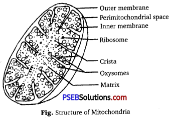
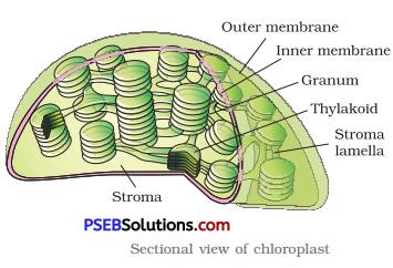
Chloroplasts and mitochondria are double membrane bound organelles.
Characteristics of Mitochondria
(i) The mitochondria are sausage-shaped or cylindrical having a diameter of 0.2-1.0 pm and average 0.5 pm and length 1.0-4.1 μm.

(ii) Each mitochondrion is a double membrane bound structure.

(iii) The inner compartment is called the matrix. The outer membrane of mitochondria forms the continuous limiting boundary of the organelle.

(iv) The inner membrane forms a number of infoldings called the cristae (single crista) towards the matrix. The cristae increase the surface area.

(v) The two membranes have their own specific enzymes associated with the mitochondrial function. The matrix of mitochondria also possess single circular DNA molecule, a few RNA molecules, ribosomes (70 s) and the components required for the synthesis of proteins.

Functions of Mitochondira: Mitochondria are the sites of aerobic respiration. They produce cellular energy in the form of ATP hence they are known as power house of the cell.
PSEB 11th Class Biology Solutions Chapter 8 Cell The Unit of Life 1
Characteristics of Chloroplasts

  • The chloroplasts are also double membrane bound organelles.
  • The space limited by the inner membrane of the chloroplast is called the stroma.

PSEB 11th Class Biology Solutions Chapter 8 Cell The Unit of Life 2

  • A number of organised flattened membranous sacs called the thylakoid are present in the stroma.
  • Thylakoids are arranged in stacks-like the piles of coins called grana.
  • In addition there are flat membranous tubules called the stroma lamellae connecting the thylakoids of the different grana.
  • The stroma of the thylakoids enclose a space called a lumen.
  • The stroma of the chloroplast contains enzymes required for the synthesis of carbohydrates and proteins.
  • Chlorophyll pigments are present in the thylakoids.
  • The ribosomes of the chloroplants are smaller (70S) than the cytoplasmic ribosomes (80S).

Functions of chloroplasts: The chloroplasts contain chlorophyll and carotenoid pigments which are responsible for trapping light energy essential for photosynthesis.

Question 8.
What are the characteristics of prokaryotic cells?
Answer:
Characteristics of Prokaryotic Cells

  • A prokaryotic cell, i.e., of bacteria is surrounded by a cell membrane. The cell wall in turn is surrounded by a slimy layer.
  • Absence of well organised chloroplast, mitochondria and nucleus.
  • The true nucleus with nuclear membrane, nucleolus is absent. It is known as nucleoid. The DNA of a prokaryotic cell is circular and not associated with basic proteins.
  • The cytoplasm is filled with dense granules. Most of these granules are ribosomes.
  • In chloroplast the scattered thylakoids are present. They are not organised in the form of stacks.

PSEB 11th Class Biology Solutions Chapter 8 Cell: The Unit of Life

Question 9.
Multicellular organisms have division of labour. Explain.
Answer:
The body of a multicellular organism has cell as a basic structural unit. The cells organised to form tissues such as blood, bone, etc. The tissues organised to form organs such as heart, kidney, etc. The organs then organised to form organ systems such as digestive system, reproductive system and respiratory system, etc. The various organ systems of organism get arranged to form a complete individual.

Question 10.
Cell is the basic unit of life. Discuss in brief.
Answer:
All organisms begin their life in a single cell. Certain organisms complete their life cycle as a single cell. They are called unicellular or acellular organisms, e.g., Amoeba, Chlamydomonas, bacteria and yeast. In other organisms, the, single cell undergoes divisions to form multicellular body. Body of human being, is made up of trillion of cells. All the cells of an organism carry the same genetic material, develop from same pre-existing cells and possess several organelles to perform various life activities. The cells are therefore, basic unit of life and structural unit of an organism.

Question 11.
What are nuclear pores? State their function.
Answer:
Nuclear pores are. small apertures present in the nuclear membrane.
Functions of Nuclear Pores: Nuclear pores are highly selective in their permeation. They allow outward passage of newly formed ribosome units but prevent the entry of active ribosomes. Proteins synthesised in the cytoplasm enter the nuclear through nuclear pores but ions like K+ Na+ or Cl may not be able to gain entrance.

Question 12.
Both lysosomes and vacuoles are endomembrane structures, yet they differ in terms of their functions. Comment.
Answer:
Both lysosomes and vacuoles are covered by a single membrane. Both of them perform different types of functions. Lysosomes contain hydrolysing enzymes and can hydrolyse all types of organic substances, – except cellulose. They perform phagocytic function. Therefore, they are known as suicidal bags.

The vacuoles are non-cytoplasmic sacs which, are also covered by a membrane. The sap vacuoles store sap or water with dissolved organic and inorganic substances. They maintain osmotic pressure or turgidity. Some freshwater invertebrates such as Amoeba, Paramecium occur contractile vacuoles, which perform osmoregulation and excretion. There is another type of vacuoles such as food vacuole which store food and gas vacuoles which store metabolic gases and take part in buoyancy regulation.

Thus, both lysosomes and vacuoles differ from each other in the type of functions they perform.

PSEB 11th Class Biology Solutions Chapter 8 Cell: The Unit of Life

Question 13.
Describe the structure of the following with the help of labelled diagrams:
(i) Nucleus
(ii) Centrosome
Answer:
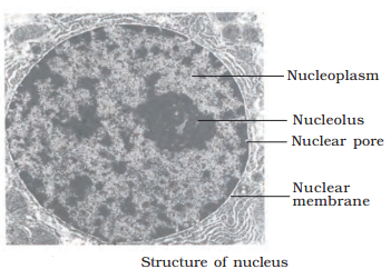
(i) Nucleus: It is a double membrane bounded protoplasmic body that carries hereditary information. Chemically it contains DNA, basic proteins, non-basic proteins, RNA, lipids and minerals, etc.
(a) Nuclear envelope: It is made up of two nuclear membranes separated by 10-70 nm perinuclear space. The outer membrane is rough due to the presence of ribosomes. Nuclear envelope has many pores with diameter
200-800 Å.

(b) Nucleoplasm or nuclear matrix: It is a colloidal complex that fills the nucleus. Nucleoplasm contains raw material for synthesis of DNA and RNA

(c) Chromatin: It is a fibrous hereditary material formed by DNA-histone complex. Some non-histone proteins and also RNA.- A single human cell has about 2 metre long thread of DNA distributed among its 46 chromosomes.

(d) Nucleolus: It was originally discovered by Fontana (1781) and given the present name by Bowman (1840). It is naked roughly rounded darkly stained structure that ‘ is attached to chromatin at specific spot called Nucleolar Organiser Region (NOR). Nucleolus is the site for eLaboration of rRNA and synthesis of ribosomes. It is therefore, known as ribosomal factory.
PSEB 11th Class Biology Solutions Chapter 8 Cell The Unit of Life 3

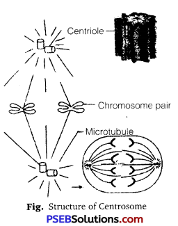
Centrosome: It is an organelle usually containing two cylindrical structure called centrioles. They are surrounded by amorphous pericentriolar materials. Both the centrioles in a centrosome lie perpendicular to each other in which
each his an organisation like the cartwheel.

They are made up of nine evenly spaced peripheral fibrils of tubulin protein the central part of the proximal region of centriole is called hub, which is connected with tubules of the peripheral triplet by radial spokes made of protein. The centriole form the basal bodies of cilia or flagella and spindle fibres that give rise to spindle apparatus during cell division in animal life.
PSEB 11th Class Biology Solutions Chapter 8 Cell The Unit of Life 4

PSEB 11th Class Biology Solutions Chapter 8 Cell: The Unit of Life

Question 14.
What is a centromere? How does the position of the centromere form the basis of classification of chromosomes? Support your answer with a diagram showing the position of centromere on different types of chromosomes.
Answer:
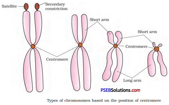
Centromere is a narrow non-stainable area, which join two similar threads or chromatids of a late prophase or metaphase chromosome. The two parts of the chromosome on either side of the centromere are known as arm. They may be isobranchial (equal) or heterobranchial (unequal in length). Depending upon the position of the centromere, the chromosomes are classified as follows:

  • Acrocentric Chromosome: The centromere is sub-terminal, at anaphasic stage appear J-shaped.
  • Sub-metacentric Chromosome: The centromere is sub-median and the anaphasic chromosome appear L-shaped.
  • Metacentric Chromosome: The centromere is in the middle and the chromosome appears V-shaped in anaphase.
  • Telocentric Chromosome: Centromere is terminal, anaphasic stage is I-shaped.

PSEB 11th Class Biology Solutions Chapter 8 Cell The Unit of Life 5
Depending upon the number of centromeres a chromosome possess, it may be monocentric, dicentric (two centromeres), polycentric (many centromeres), acentric chromosome (having no centromere).

PSEB 11th Class Biology Solutions Chapter 7 Structural Organisation in Animals

Punjab State Board PSEB 11th Class Biology Book Solutions Chapter 7 Structural Organisation in Animals Textbook Exercise Questions and Answers.

PSEB Solutions for Class 11 Biology Chapter 7 Structural Organisation in Animals

PSEB 11th Class Biology Guide Structural Organisation in Animals Textbook Questions and Answers

Question 1.
Answer in one word or one line:
(i) Give the common name of Periplanata americana.
(ii) How many spermathecae are found in earthworm?
(iii) What is the position of ovaries in cockroach?
(iv) How many segments are present in the abdomen of cockroach?
(v) Where do you find Malpighian tubules?
Answer:
(i) American cockroach
(ii) 4 pairs
(iii) Under the 4th, 6th abdominal terga
(iv) 10 segments in adults
(v) At the junction of midgut and hindgut in cockroach.

Question 2.
Answer the following:
(a) What is the function of nephridia?
(b) How many types of nephridia are found in earthworm based on their location?
Answer:
(a) The Function of Nephridia: The nephridia regulate the volume and composition of the body fluids. The nephridium starts out as a funnel that collects excess fluid from coelomic chamber. The funnel connects with a tubular parts of the nephridium, which delivers the wastes through a pore to the surface in the body wall in the digestive tube.

(b) Based on their location, there are following three types of nephridia in earthworm:

  • Septal nephridia.
  • Pharyngeal nephridia.
  • Integumentary nephridia.

PSEB 11th Class Biology Solutions Chapter 7 Structural Organisation in Animals

Question 3.
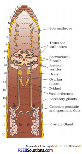
Draw a labelled diagram of the reproductive organs of an earthworm.
Answer:
PSEB 11th Class Biology Solutions Chapter 7 Structural Organisation in Animals 1

Question 4.
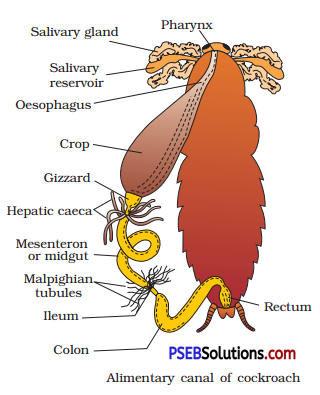
Draw a labelled diagram of alimentary canal of a cockroach.
Answer:
PSEB 11th Class Biology Solutions Chapter 7 Structural Organisation in Animals 2

Question 5.
Distinguish between the following:
(a) Prostomium and peristomium.
(b) Septal nephridium and pharyngeal nephridium.
Answer:
(a) Prostomium and Peristomium: The first segment of earthworm with a ventral mouth is known as peristomium. Prostomium is a dorsal, lobe which is present on the ventral mouth.

(b) Septal Nephridia and Pharyngeal Nephridia: Septal nephridia are present on both the sides of intersegmental septa of segment 15 to the ‘ last that open into intestine.
The pharyngeal nephridia are closed (no nephrostome) nephridia present as three paired groups (of about 100) in 4th, 5th and 6th segments.

PSEB 11th Class Biology Solutions Chapter 7 Structural Organisation in Animals

Question 6.
What are the cellular components of blood?
Answer:
The cellular components of blood are red blood cells, white blood cells and platelets.

Question 7.
What are the following and where do you find them in animal body (a) Chondrocytes (b) Axons (c) Ciliated epithelium.
Answer:
(a) Chondrocytes: These are the matrix secreting cells of the cartilage. These are found in the cartilage of connecting tissue.

(b) Axon: It is a long fibre, the distal end of which is branched. Each branch terminates as a bulb like structure called synaptic knob. The axon transmit nerve impulses away from the cell body.

(c) Ciliated Epithelium: If the columnar or cuboidal cells of columnar and cuboidal epithelium bear cilia on their free surface they are called ciliated epithelium.

Question 8.
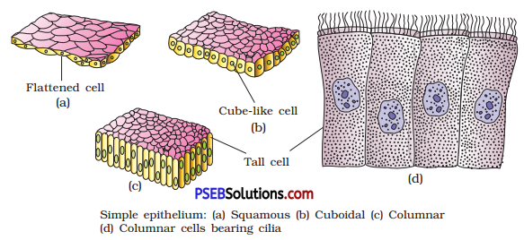
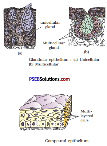
Describe various types of epithelial tissues with the help of labelled diagrams.
Answer:
Epithelial Tissues: Epithelial tissues provide covering to the inner and outer lining of various organs. There are following two types of epithelial tissues :
1. Simple epithelium: Simple epithelium is composed of a single layer of cells. It functions as a lining for body cavities, ducts and tubes.

2. Compound epithelium: The compound epithelium consists of two or more cell layers. It has protective function as it does in our skin. It covers the dry surface of the skin, the moist surface of buccal cavity, pharynx, inner lining of ducts of salivary glands and of pancreatic ducts.
On the basis of structural modifications of the cells, simple epithelium is further divided into three types. These are :
(a) Squamous epithelium: The squamous epithelium is made of a single thin layer of flattened cells with irregular boundaries. They are found in the walls of blood vessels and air sacs of lungs and are involved in functions like forming a diffusion boundary.

(b) Cuboidal epithelium: The cuboidal epithelium is composed of a single layer of cube-like cells. This is commonly found in the ducts of glands and tubular parts of nephrons in kidneys. Its main functions are secretion and absorption.

(c) Columnar epithelium: The columnar epithelium is composed of a single layer of tall and slender cells. They are found in the lining of stomach and intestine and help in absorption and secretion.
(i) When the columnar or cuboidal cells bear cilia on their free surface they are called ciliated epithelium. Their function is to move particles or mucus in a specific direction over the epithelium organs like bronchioles and fallopian tubules.
(ii) Some of the columnar or cuboidal cells get specialized for secretion are called glandular epithelium. They are unicellular and multicellular.
PSEB 11th Class Biology Solutions Chapter 7 Structural Organisation in Animals 3
PSEB 11th Class Biology Solutions Chapter 7 Structural Organisation in Animals 4

PSEB 11th Class Biology Solutions Chapter 7 Structural Organisation in Animals

Question 9.
Distinguish between:
(a) Simple epithelium and compound epithelium
(b) Cardiac muscle and striated muscle
(c) Dense regular and dense irregular connective tissue
(d) Adipose tissue and blood tissue
(e) Simple gland and compound gland
Answer:
(a) Differences between Simple and Compound Epithelium

Simple Epithelium Compound Epithelium
1. It is composed of a single layer of It consists of two or more cell layers.
2. It functions as a lining for body cavities, ducts and tubes. It is protective in function like our skin.

(b) Differences between Cardiac and Striated Muscle

Cardiac Muscle Striated Muscle
1. It occurs only in the wall of heart. It occurs in the body wall, limb, tongue, pharynx, etc.
2. They are short and cylindrical with truncate ends. They are long and cylindrical with blunt ends.
3. They have nerve supply from brain and autonomous nerve system They have nerve supply from central nervous system.

(c) Differences between Dense Regular and Dense Irregular Connective Tissues

Dense Regular Connective Tissue Dense Irregular Connective Tissue
Collagen fibres are present in rows between many parallel bundle of fibres.
Examble: Tendons
Fibroblasts and many fibre are present that are oriented differently.
Examble: Cartilage, bones and blood.

(d) Differences between Adipose Tissue and Blood Tissue

Adipose Tissue Blood Tissue
1. It is a soft gel like connective tissue. It is a fluid connective tissue.
2. It is partitioned into lobules by septa. There are no partitions.
3. It is a storage tissue. It is a transport tissue.
4. Matrix is secreted by the cells. Matrix is not secreted by the cells.
5. It contain fibres. Fibres are not conspicuous.
6. Adipocytes contain fat droplets. No cell of the tissue contains fat droplets.

(e) Differences between Simple and Compound Gland

Simple Gland Compound Gland
1. These glands have single unbranched duct. These glands have branched system of ducts.
2. These may be simple tubular glands, simple coiled tubular glands and simple alveolar glands. These may be compound tubular glands, compound alveolar glands

PSEB 11th Class Biology Solutions Chapter 7 Structural Organisation in Animals

Question 10.
Mark the odd one in each series:
(a) Areolar tissue, blood, neuron, tendon
(b) RBC, WBC, platelets, cartilage
(c) Exocrine, endocrine, salivary gland; ligament
(d) Maxilla, mandible, labrum, antennae
(e) Protonema, mesothorax, metathorax, coxa
Answer:
(a) Neuron,
(b) Cartilage,
(c) Ligament,
(d) Antennae,
(e) Protonema.

Question 11.
Match the terms in column I with those in column II.

Column I Column II
A. Compound epithelium 1. Alimentary canal
B. Compound eye 2. Cockroach
C. Septal nephridia 3. Skin
D. Open circulatory system 4. Mosaic vision
E. Typhlosole 5. Earthworm
F. Osteocytes 6. Phallomere
G. Genitalia 7. Bone

Answer:

Column I Column II
A. Compound epithelium 3. Skin
B. Compound eye 4. Mosaic vision
C. Septal nephridia 5. Earthworm
D. Open circulatory system 2. Cockroach
E. Typhlosole 1. Alimentary canal
F. Osteocytes 7. Bone
G. Genitalia 6. Phallomere

Question 12.
Mention briefly about the circulatory system of earthworm.
Answer:
Pheretima exhibits a closed type of blood vascular system, consisting of blood vessels, capillaries and heart. Due to closed circulatory system, blood is confined to the heart and blood vessels. Contractions keep blood circulating in one direction. Smaller blood vessels supply the gut, nerve cord, and the body wall. Blood glands are present on the 4th, 5th and 6th segments. They produce blood cells and haemoglobin which is dissolved in blood plasma. Blood cells are phagocytic in nature. Earthworms lack specialised breathing devices. Respiratory exchange occurs through moist body surface into their blood stream.

PSEB 11th Class Biology Solutions Chapter 7 Structural Organisation in Animals

Question 13.
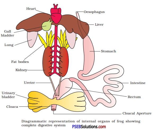
Draw a neat diagram of digestive system of frog.
Answer:
PSEB 11th Class Biology Solutions Chapter 7 Structural Organisation in Animals 5

Question 14.
Mention the function of the following:
(a) Ureters in frog
(b) Malpighian tubules
(c) Body wall in earthworm
Answer:
(a) Functions of Ureters in Frog: In male frog, two ureters emerge from the kidneys. The ureters act as urinogenital duct which opens into the cloaca. Thus, the ureters carry both sperms and excretory wastes to the cloaca. In female frog, the ureters and oviduct open separately in the cloaca. The ureters in frog, thus acts as carrier of sperms and ova.

(b) Functions of Malpighian Tubules of Cockroach: Excretion is carried out by Malpighian tubules. Each tubule is lined by glandular cells. They absorb excretory waste products and converts them into uric acid which is excreted out through the hindgut.

(c) Functions of Body Wall of Earthworm: The body wall of earthworm has five layers – cuticle, epidermis, circular muscle layer, longitudinal muscle layer, peritoneum.

  • Cuticle is a non-cellular elastic layer.
  • The columnar cells of body wall provide support and therefore, are also known as supporting cells.
  • Epidermis also has gland cells, receptor cells and basal cells.
  • The glandular cell secrete mucus and thus, keep the skin moist, this
    help in cutaneous respiration.
  • The last layer of the body wall is the outer membrane of the coelom called coelomic epithelium. The various muscle layers of the body wall provide strength and rigidity.

PSEB 11th Class Biology Solutions Chapter 16 Digestion and Absorption

Punjab State Board PSEB 11th Class Biology Book Solutions Chapter 16 Digestion and Absorption Textbook Exercise Questions and Answers.

PSEB Solutions for Class 11 Biology Chapter 16 Digestion and Absorption

PSEB 11th Class Biology Guide Digestion and Absorption Textbook Questions and Answers

Question 1.
Choose the correct answer among the following:
(i) Gastric juice contains
(a) pepsin, lipase and rennin
(b) trypsin, lipase and rennin
(c) trypsin, pepsin and lipase
(d) trypsin, pepsin and rennin

(ii) Succus entericus is the name given to
(a) a junction between ileum and large intestine
(b) intestinal juice
(c) swelling in the gut
(d) appendix
Answer:
(i) (a) Pepsin, lipase, and rennin
(ii) (b) Intestinal juice.

Question 2.
Match column I with column II.

Column I Column II
A. Bilirubin and biliverdin 1. Parotid
B. Hydrolysis of starch 2. Bile
C. Digestion of fat 3. Lipases
D. Salivary gland 4. Amylases

Answer:

Column I Column II
A. Bilirubin and biliverdin 2. Bile
B. Hydrolysis of starch 4. Amylases
C. Digestion of fat 3. Lipases
D. Salivary gland 1. Parotid

PSEB 11th Class Biology Solutions Chapter 16 Digestion and Absorption

Question 3.
Answer briefly:
(a) Why are villi present in the intestine and not in the stomach?
(b) How does pepsinogen change into its active form?
(c) What are the basic layers of the wall of alimentary canal?
(d) How does bile help in the digestion of fats?
Answer:
(a) The mucosa layer of alimentary canal forms small finger-like foldings called villi in the small intestine. The cells lining the villi produce numerous microscopic projections called microvilli giving a brush border appearance. These modifications increase the surface area enormously.
Villi are supplied with the network of capillaries and large lymph vessel called the lacteal mucosal.

(b) The inactive form of enzyme pepsinogen is activated by Rd.

(c) The wall of alimentary canal from esophagus to rectum possesses four layers namely serosa, muscularis, sub-mucosa and mucosa. Serosa is the outermost layer, followed by muscularis, sub-mucosa and mucosa.

(d) Bile salts help in emulsification of lipids and activate the lipases.

Question 4.
State the role of pancreatic juice in digestion of proteins.
Answer:
The pancreatic juice contains inactive enzymes trypsinogen, chymotrypsinogen, procarboxypeptidases, amylases, lipases, and nucleases. Trypsinogen is aëtivated by an enzyme, enterokinase, secreted by the intestinal mucosa into active trypsin, which in turn activates the other enzymes in the pancreatic juice. Proteins, proteases and peptones (partially hydrolyzed proteins) in the chyme reaching the intestine are acted upon by the proteolytic enzymes of pancreatic juice as given below:
PSEB 11th Class Biology Solutions Chapter 16 Digestion and Absorption 1

Question 5.
Describe the process of digestion of protein in stomach.
Answer:
The food mixes thoroughly with the acidic gastric juice of the stomach by the churning movements of its muscular wall and is called the chyme. The pepsinogen, on exposure to hydrochloric acid gets converted into the active enzyme pepsin, the proteolytic enzyme of the stomach. Pepsin converts proteins into proteoses and peptones (peptides).
PSEB 11th Class Biology Solutions Chapter 16 Digestion and Absorption 2
HCl provides the acidic pH (pH 1.8) optimal for pepsins. Rennin is a proteolytic enzyme found in gastric juice of infants which helps in the digestion of milk proteins.

Question 6.
Give the dental formula of human beings.
Answer:
The dental formula of human beings is
\(\frac{2123}{2123} \times 2\).

PSEB 11th Class Biology Solutions Chapter 16 Digestion and Absorption

Question 7.
Bile juice contains no digestive enzymes, yet it is important for digestion. Why?
Answer:
Bile is yellowish-green alkaline solution with 89-98% water, having no digestive enzymes. The bile released into the duodenum contains bile pigments (bilirubin and biliverdin), bile salts, cholesterol, and phospholipids but no enzymes. Bile helps in emulsification of fats, i.e., breaking down of the fats into very small micelles. Bile also activates lipases.

Question 8.
Describe the digestive role of chymotrypsin. Which two other digestive enzymes of the same category are secreted by its source gland?
Answer:
Chymotrypsin is the active form of chymotrypsinogen. It is activated by trypsin. It curdles milk. Nucleases like DNA ase and RNAase and pancreatic lipase are other enzymes secreted by the pancreas.

Question 9.
How are polysaccharides and disaccharides digested?
Answer:
The chemical process of digestion of carbohydrates is initiated in the oral cavity by the hydrolytic action of the carbohydrate splitting enzyme, the salivary amylase. About 30 percent of starch is hydrolyzed here by this enzyme (optimum pH 6.8) into a disaccharide-maltose. Further, carbohydrates in the chyme are hydrolyzed by pancreatic amylase into disaccharides.
PSEB 11th Class Biology Solutions Chapter 16 Digestion and Absorption 3

Question 10.
What would happen if HCl were not secreted in the stomach?
Answer:
The mucus and bicarbonates present in the gastric juice play an important role in lubrication and protection of the mucosal epithelium from excoriation by the highly concentrated hydrochloric acid.
PSEB 11th Class Biology Solutions Chapter 16 Digestion and Absorption 4
HCl provides the acidic pH (pH 1.8) optimal for pepsins. Rennin is a proteolytic enzyme found in gastric juice of infants which helps in the digestion of milk proteins. Small amount of lipases are also secreted by gastric glands.

PSEB 11th Class Biology Solutions Chapter 16 Digestion and Absorption

Question 11.
How does butter in your food get digested and absorbed in the body?
Answer:
Bile helps in emulsification of fats, i. e., breaking down of the fats into very small micelles. Bile also activates lipases.

Question 12.
Discuss the main steps in the digestion of proteins as the food passes through different parts of the alimentary canal.
Answer:
Digestion of Protein in Stomach: The proenzyme pepsinogen, on exposure to HCl, gets converted into active enzyme pepsin.
PSEB 11th Class Biology Solutions Chapter 16 Digestion and Absorption 5
Pepsin always outs in acidic medium (pH 1.8). In infants, main proteins are digested by rennin.

Digestion of Protein in Small Intestine: Pancreatic juice contains proenzyme trypsinogen. It is activated by enterokinase, secreted by intestinal mucosa, into active trypsin. Trypsin acts in alkaline medium.
PSEB 11th Class Biology Solutions Chapter 16 Digestion and Absorption 6
The dipeptides are changed into amino acids by the enzyme succus enterics (intestinal juice).
PSEB 11th Class Biology Solutions Chapter 16 Digestion and Absorption 7

Question 13.
Explain the term ‘the codont’ and ‘diphyodont’.
Answer:
Each tooth is embedded in a socket of jaw bone. This type of attachment is called thecodont. The majority of mammals including human beings forms two sets of teeth during their life, a set of temporary milk or deciduous teeth replaced by a set of permanent or adult teeth. This type of dentition is called diphyodont.

Question 14.
Name different types of teeth and their number in an adult human.
Answer:
An adult human has 32 permanent teeth, which are of four different types (heterodont dentition), i.e., incisors (I), canine (C), premolars (Pm), and molars (M), and their number are 4, 2, 4, 6 respectively.

PSEB 11th Class Biology Solutions Chapter 16 Digestion and Absorption

Question 15.
What are the functions of liver?
Answer:
Liver is the largest gland in human body which is mainly responsible for the digestion of food.
Role of liver in digestion of food :

  • Its hepatic cells secrete bile juice which passes through the hepatic duct into the gall bladder.
  • It has its major role in digestion and processing of proteins.
  • Bile secreted by it is mainly responsible for digestion of fats for easy absorption in the body.
  • It also responsible for the removal of toxins from blood.

PSEB 11th Class Biology Solutions Chapter 6 Anatomy of Flowering Plants

Punjab State Board PSEB 11th Class Biology Book Solutions Chapter 6 Anatomy of Flowering Plants Textbook Exercise Questions and Answers.

PSEB Solutions for Class 11 Biology Chapter 6 Anatomy of Flowering Plants

PSEB 11th Class Biology Guide Anatomy of Flowering Plants Textbook Questions and Answers

Question 1.
State the location and function of different types of meristems.
Answer:
A meristematic tissue represents a group of cells that have retained the power of division throughout the life of an individual. The meristematic tissues are of three types, i.e., apical, intercalary and lateral meristem.

  • Apical Meristem: These meristems are present at the apices of shoot and roots of the plants. Apical meristems are responsible for the increase in length of all the primary tissues.
  • Intercalary Meristem: It is the meristem that occurs between the mature tissues. They occur in grasses and regenerate parts removed by the grazing herbivores. It contributes to the formation of the primary plant body.
  • Lateral Meristem: It occurs in the mature regions of roots arid shoots of many plants, particularly that produce woody axis. These meristems are responsible for producing the secondary tissues.

Question 2.
Cork cambium forms tissues that form the cork. Do you agree with this statement? Explain.
Answer:
Yes, cork cambium forms tissues that form cork. As the stem continues to increase in girth another meristematic tissue called cork cambium or phellogen develops in cortex region of stem. The phellogen cuts off cells on both sides. The outer cells differentiate into cork or phellem. The inner cells differentiate into secondary cortex or phelloderm. Cork is impervious to water due to suberin and provides protection to underlying tissues.

PSEB 11th Class Biology Solutions Chapter 6 Anatomy of Flowering Plants

Question 3.
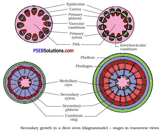
Explain the process of secondary growth in the stems of woody angiosperms with the help of schematic diagrams. What is its significance?
Answer:
Secondary growth in stems of woody angiosperms occur by two types of cambia, i.e., vascular cambium and cork cambium.
(i) Vascular Cambium: Certain cells of medullary rays become meristematic to form interfascicular cambium. The fascicular cambium and the interfascicular cambium join to form a complete ring called cambial ring. The cells of the cambial ring undergo mitotic divisions and produce secondary phloem on its outer side and secondary xylem on its inner side.

At places, vascular cambium possesses ray initials. They form vascular rays, phloem rays in secondary phloem and wood rays in secondary xylem.

As new secondary phloem becomes functional, the previous older phloem gets crushed. Secondary xylem or wood persists. As a result wood grows

with age in the form of annual rings. In each annual ring, there is wide or broader spring or early wood or spring wood and narrow autumn or late wood.

In old stems, the central part of wood becomes non-functional and dark coloured due to tyloses and deposit of resins, gums, tannins. It is called r duramen or heartwood. The outer, functional wood is called sapwood.

(ii) Cork Cambium: As the stem continues to increase in girth due to the activity of vascular cambium the outer cortical and epidermal layers get broken and need to be replaced to provide new protective cell layers. In this way, cork cambium or phellogen develops in the cortex region. Phellogen cuts of cells on both sides.

The outer cells differentiate into cork or phellem while, the inner cells ; differentiate into secondary cortex or phelloderm. Due to the activity of cork cambium, pressure builds up on remaining layers peripheral to
phellogen and ultimately these layers die and slough off. At places, aerating pores called lenticels develop, which have loosely arranged , complementary cells.
PSEB 11th Class Biology Solutions Chapter 6 Anatomy of Flowering Plants 1
Significance of Secondary Growth

  • It replaces old non-functional tissues.
  • It provides fire proof, insect proof and insulating cover around the older plant parts.
  • Commercial cork is a product of secondary growth.
  • Wood is the product of secondary growth.

PSEB 11th Class Biology Solutions Chapter 6 Anatomy of Flowering Plants

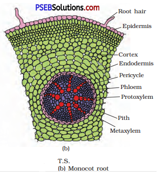
Question 4.
Draw illustrations to bring out the anatomical difference between:
(a) Monocot root and Dicot root
(b) Monocot stem and Dicot stem
Answer:
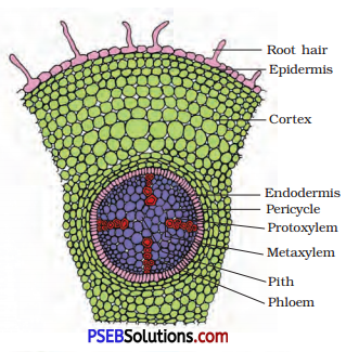
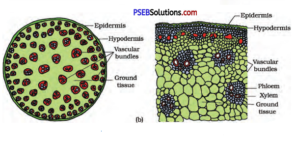
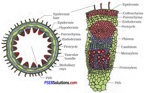
PSEB 11th Class Biology Solutions Chapter 6 Anatomy of Flowering Plants 2
(a) Anatomical Differences between Monocot Root and Dicot Root
(i) Anatomy of Monocot Root
(a) The structure of epidermis, cortex, endodermis and pericycle of a monocot root resembles exactly those of a dicot root.
(b) Vascular bundles are radial, and polyarch.
(c) Pith is large and well-developed; it is composed of parenchyma cells

(ii) Anatomy of dicot root
PSEB 11th Class Biology Solutions Chapter 6 Anatomy of Flowering Plants 3
T.S of Dicot root
(a) Epidermis is single layered and many cells bear root hairs; cuticle is absent.
(b) Cortex is made of several layers of parenchyma cells.
(c) Endodermis consists of a single-layer of cells. The cells have a deposition of suberin, in the form of casparian strips, on their radial and tangential walls.
(d) Pericycle comprises a few layers of specialised parenchyma cells inner to the endodermis.
(e) Vascular bundles are radial and may range between two and six, though commonly there are four groups; i.e., tetrarch; xylem is exarch.
(f) Pith is very small and made of parenchyma cells.

(b) Anatomical Differences between Monocot Stem and Dicot stem
(i) Anatomy of monocot stem
(a) Epidermis is single layered and trichomes are absent; cuticle is present on its outer surface.
(b) Hypodermis consists of two or three layers of sclerenchyma cells.
(c) Ground tissue is parenchymatous and is not differentiated into cortex or pith.
(d) Vascular bundles are many and scattered in the ground tissue; they vary in size.
PSEB 11th Class Biology Solutions Chapter 6 Anatomy of Flowering Plants 4
T.S. of Monocot Stem

  • Each vascular bundle is surrounded by sclerenchymatous bundle sheath.
  • The vascular bundles are conjoint and closed; xylem is endarch and characteristically a protoxylem lacuna is present.

(ii) Anatomy of dicot stem
(a) Epidermis is the outermost layer of cells; externally it is covered with a cuticle and may bear trichomes and a few stomata.
(b) Hypodermis consists of a few layers of collenchyma cells, just below the epidermis.
(c) Cortex consists of parenchyma cells.
(d) Endodermis is single layered and the cells are rich in starch grains and hence it is also referred to as starch sheath.
(e) Pericyde occurs inner to the endodermis, above the phloem of vascular bundles in the form of semi-lunar patches (hence also referred to as bundle caps); it is composed of sclerenchyma.
T.S. of Dicot Stem
PSEB 11th Class Biology Solutions Chapter 6 Anatomy of Flowering Plants 5
(f) Vascular bundles are characteristically arranged in the form of a ring.

  • Each vascular bundle is conjoint and open with intra-fascicular cambium; xylem is endarch.
    (g) Medullary rays are the few layers of radially placed parenchyma cells, in between the vascular bundles.
    (h) Pith is composed of parenchyma cells.

PSEB 11th Class Biology Solutions Chapter 6 Anatomy of Flowering Plants

Question 5.
Cut a transverse section of young stem of a plant from your school garden and observe it under the microscope. How would you ascertain whether it is a monocot stem or a dicot stem? Give reasons.
Answer:
Transverse section of a monocot stem possess following characters:

  • Dumbbell-shaped guard cells in stomata in epidermis.
  • Sclerenchymatous hypodermis.
  • No concentric arrangement of internal tissues.
  • Unifrom ground tissue showing no tissue differentiation.
  • More than 8 scattered vascular bundles.
  • Bundle sheath is present.
  • No secondary growth normally.
  • Xylem vessels arranged in Y-shaped manner.
  • Protoxylem cavity usually present in vascular tissues.

Transverse section of a dicot stem possess following characters:

  • Kidney-shaped guard cells in stomata present in epidermis.
  • Collenchymatous hypodermis.
  • Concentric arrangement of internal tissues.
  • Differentiation of ground tissue into cortex, endodermis, pericycle and pith.
  • The vascular bundles are arranged in a ring.
  • Conjoint, collateral and open vascular bundles.
  • Without bundle sheath.
  • Secondary growth takes place.
  • Xylem vessels arranged in rows.

[Note: For figures refer to Q.No. 4]

Question 6.
The transverse section of a plant material shows the following anatomical features:
(a) The vascular bundles are conjoint, scattered and surrounded by a sclerenchymatous bundle sheaths.
(b) Phloem parenchyma is absent. What will you identify it as?
Answer:
It is a transverse section of monocotyledonous stem.

Question 7.
Why are xylem and phloem called complex tissues?
Answer:
Xylem and phloem are made up of more than one type of cells that is why they are called as complex tissues :
(i) Xylem is composed of four different kinds of elements, namely-tracheids, vessels, xylem fibres and xylem parenchyma. Tracheids are dead tube-like cells which are thick walled, vessels are made up of large number of tube cells placed end to end. Xylem fibres are thick walled cells that maybe septate and aseptate. Xylem parenchyma is living and thin walled cells.

(ii) Phloem is composed of sieve tube elements, companion cells, phloem parenchyma and phloem fibres. Sieve tube elements are tube-like cells, whereas phloem parenchyma are living cells and phloem fibres are thick walled lignified cells.

PSEB 11th Class Biology Solutions Chapter 6 Anatomy of Flowering Plants

Question 8.
What is stomatal apparatus? Explain the structure of stomata with a labelled diagram.
Answer:
The minute pores present in the epidermis are known as stomata. The stomata may be surrounded by either bean-shaped (in dicots) or by dumb-bell-shaped (in monocots) guard cells. The guard cells in turn are surrounded by other epidermal cells, which are known as subsidiary or
PSEB 11th Class Biology Solutions Chapter 6 Anatomy of Flowering Plants 6
accessory cells. The stomatal aperture, guard cells, accessory cells constitute the stomatal apparatus.

Question 9.
Name the three basic tissue systems in the flowering plants. Give the tissue names under each system.
Answer:
The three basic; tissue systems in flowering plants are as follows:

  • Epidermal Tissue System: The tissue related to this system are epidermis, cuticle and wax, stomata and trichomes.
  • Ground Tissue System: It consists of cortex, endodermis, pericycle, medullary rays, pith and ground tissue of leaves.
  • Vascular Tissue System: It contains conducting tissues like xylem and phloem.

Question 10.
How is the study of plant anatomy useful to us?
Answer:
Plant anatomy is the study of internal structure of living organisms.

  • It describes the tissues involved in assimilation of food and its storage, transportation of water, i.e., xylem tissue, transportation of minerals. i.e., phloem and those involved in providing mechanical support to the plant,
  • Study of internal structure of plants helps to understand their adaptations of diverse environments.
  • The study of plant anatomy also help in understanding the functional organisation of higher plants.

Question 11.
What is periderm? How does periderm formation take place in
the dicot stems?
Answer:
Phellogen, phellem and phelloderm are collectively called as periderm. Phellogen develops usually in the cortex region. Phellogen is a couple of layers thick. Phellogen cuts off cells on both sides. The outer cells 1 differentiate into cork or phellem, while the inner cells differentiate into secondary cortex or phelloderm. All these together form periderm.

PSEB 11th Class Biology Solutions Chapter 6 Anatomy of Flowering Plants

Question 12.
Describe the internal structure of a dorsiventral leaf with the help of labelled diagrams.
Answer:
The vertical section of a dorsiventral leaf through the lamina shows three ; main parts namely, epidermis, mesophyll and vascular system. The epidermis which covers both the upper surface (adaxial epidermis) and lower surface (abaxial epidermis) of the leaf has a conspicuous cuticle. The abaxial epidermis generally bears more stomata than the adaxial epidermis. The latter may even lack stomata. The tissue between the upper and the lower epidermis is called the mesophyll. Mesophyll, which possesses chloroplasts and carry out photosynthesis, is made up of parenchyma. It has two types of cells – the palisade parenchyma and the spongy parenchyma.

The adaxially placed palisade parenchyma is made up of elongated cells, which are arranged vertically and parallel to each other. The oval or round and loosely arranged spongy parenchyma is situated below the palisade cells and extends to the lower epidermis. There are numerous large spaces and air cavities between these cells. Vascular system includes vascular bundles, which can be seen in the veins and the midrib. The size of the vascular bundles are dependent on the size of the veins. The veins vary in thickness in the reticulate venation of the dicot leaves. The vascular bundles are surrounded by a layer of thick walled bundle sheath cells.
T.S. of Dorsiventral Leaf
PSEB 11th Class Biology Solutions Chapter 6 Anatomy of Flowering Plants 7

PSEB 11th Class Biology Solutions Chapter 17 Breathing and Exchange of Gases

Punjab State Board PSEB 11th Class Biology Book Solutions Chapter 17 Breathing and Exchange of Gases Textbook Exercise Questions and Answers.

PSEB Solutions for Class 11 Biology Chapter 17 Breathing and Exchange of Gases

PSEB 11th Class Biology Guide Breathing and Exchange of Gases Textbook Questions and Answers

Question 1.
Define vital capacity. What is its significance?
Answer:
Vital Capacity (VC): The maximum volume of air a person can breathe in after a forced expiration is called vital capacity. Vital capacity is higher in athletes and singers. Vital capacity shows the strength of our inspiration and expiration.

Question 2.
State the volume of air remaining in the lungs after a normal breathing.
Answer:
The volume of air remaining in the lungs even after a forcible expiration averages 1100 ml to 1200 ml.

Question 3.
Diffusion of gases occurs in the alveolar region only and not in the other parts of respiratory system. Why?
Answer:
Alveoli are the primaty sites of gas exchange in the respiratory system. Exchange of gases occur between blood and these tissues. O2 and CO2 are exchanged in these sites by simple diffusion mainly based on pressure/concentration gradient. The diffusion membrane for gas exchange is made up of three major layers.
These layers are :

  1. Squamous epithelium of alveoli.
  2. Endothelium of alveolar capillaries.
  3. Basement substance in between them. Its total thickness is much less than a millimetre. Therefore, all the factors in our body are favourable for diffusion of O2 from alveoli to tissues and that of CO2 from tissues to alveoli.

Question 4.
What are the major transport mechanisms for CO2? Explain.
Answer:
Transport of Carbon Dioxide: CO2 in gaseous form diffuses out of the cells into capillaries, where it is transported in following ways :
(i) Transport in dissolved form: About 7% CO2 is carried in dissolved form through the plasma because of its high solubility.
(ii) Transport as bicarbonate: The largest fraction (about 70%) is carried in plasma as bicarbonate ions (HCO3). At the tissues site, where pCO2 is high due to catabolism, CO2 diffuses into the blood (RBCs and plasma) and forms HCO3 and H.
PSEB 11th Class Biology Solutions Chapter 17 Breathing and Exchange of Gases 1
This reaction is faster in RBCs because they contain an enzyme carbonic anhydrase. Hydrogen ion released during the reaction bind to Hb, triggering the Bohr effect.
At the alveolar site, where pCO2 is low, the reaction proceeds in opposite direction forming CO2 and H2O. Thus, CO2 trapped as bicarbonate at tissue level and transported to alveoli is released as CO2.

(iii) Transport as carbaminohaemoglobin: Nearly 20-25% CO2 is carried by haemoglobin as carbaminohaemoglobin, CO2 entering the blood combines with the NH2 group of the reduced Hb.
PSEB 11th Class Biology Solutions Chapter 17 Breathing and Exchange of Gases 2
The reaction releases oxygen from oxyhaemoglobin.
Factors affecting the binding of CO2 and Hb are as follows:

  • Partial pressure of CO2.
  • Partial pressure of O2 (major factor).

In tissues, pCO2 is high and pO2 is low, more binding of CO2 occurs while, in the alveoli, pCO2 is low and pO2 is high, dissociation of CO2 from HbCO2 takes place, i.e., CO2, which is bound to Hb from the tissues is delivered at the alveoli.

PSEB 11th Class Biology Solutions Chapter 17 Breathing and Exchange of Gases

Question 5.
What will be the pO2 and pCO2, in the atmospheric air compared to those in the alveolar air?
(i) pO2 lesser and pCO2 higher
(ii) pO2 higher and pCO2 lesser
(iii) pO2 higher and pCO2 higher
(iv) pO2 lesser and pCO2 lesser
Answer:
(i) In the alveolar tissues, where low pO2, high pCO2, high H+ concentration, these conditions are favourable for dissociation of oxygen from the oxyhaemoglobin.

(ii) When there is high pO2, low pCO2, less H+ concentration and lesser temperature, the factors are all favourable for formation of oxyhaemoglobin.

(iii) When pO2 is high in the alveoli and pCO2 is high in the tissues then the oxygen diffuses into the blood and combines with oxygen forming oxyhaemoglobin and CO2 diffuses out.

(iv) When pO2 is low in the alveoli and pCO2 is low in the tissues then these conditions are favourable for dissociation of oxygen from the oxyhaemoglobin.

Question 6.
Explain the process of inspiration under normal conditions.
Answer:
Inspiration is the process during which atmospheric air is drawn in. Inspiration is initiated by the contraction of diaphragm, which increases the volume of thoracic chamber in the anteroposterior axis. The contraction of external intercostal muscles lifts up the ribs and the sternum causing an increase in the volume of the thoracic chamber in the dorso-ventral axis.

The overall increase in the thoracic volume causes a similar increase in pulmonary volume. An increase in pulmonary volume decreases the intrapulmonary pressure to less than the atmospheric pressure, which forces the air from outside to move into the lungs, i. e., inspiration. On an average, a healthy human breathes 12-16 times/minute.
PSEB 11th Class Biology Solutions Chapter 17 Breathing and Exchange of Gases 3

Question 7.
How is respiration regulated?
Answer:
Respiratory rhythm centre is primarily responsible for regulation of respiration. This centre is present in the medulla.
Pneumotaxic centre, present in the pons region, also coordinates respiration. Apart from them, receptors associated with aortic arch and carotid artery, can also recognize changes in CO2 and H+ concentration and send signal to the rhythm centre for proper action.

Question 8.
What is the effect of pCO2 on oxygen transport?
Answer:
Partial pressure of CO2 (pCO2) can interfere the binding of oxygen with haemoglobin, i.e., to form oxyhaemoglobin.
(i) In the alveoli, where there is high pO2 and low pCO2, less H+ concentration and low temperature, more formation of oxyhaemoglobin occur.

(ii) In the tissues, where low pO2, high pCO2, high H+ concentration and high temperature exist, the conditions are responsible for dissociation of oxygen from the oxyhaemoglobin.

Question 9.
What happens to the respiratory process in a man going up a hill?
Answer:
At hills, the pressure of air falls and the person cannot get enough oxygen in the lungs for diffusion in blood. Due to deficiency of oxygen, the person feels breathlessness, headache, dizziness, nausea, mental fatigue and a bluish colour on the skin, nails and lips.

PSEB 11th Class Biology Solutions Chapter 17 Breathing and Exchange of Gases

Question 10.
What is the site of gaseous exchange in an insect?
Answer:
The actual site of gaseous exchange in an insect is tracheoles and tracheolar end cells.

Question 11.
Define oxygen dissociation curve. Can you suggest any reason for its sigmoidal pattern?
Answer:
A sigmoid curve is obtained when percentage saturation of haemoglobin with O2 is plotted against the pO2.
This curve is called the oxygen dissociation curve and is highly useful in studying the effect of factors like pCO2, H+ concentration, etc., on binding of O2 with haemoglobin.
PSEB 11th Class Biology Solutions Chapter 17 Breathing and Exchange of Gases 4
In the alveoli, where there is high pO2, low pCO2, lesser H+ concentration and lower temperature, the factors are all favourable for the formation of oxyhaemoglobin, whereas in the tissues, where low pO2, high pCO2, high H+ concentration and higher temperature exist, the conditions are favourable for dissociation of oxygen from the oxyhaemoglobin. This dearly indicates that O2 gets bound to haemoglobin in the lung surface and gets dissociated at the tissues. Every 100 ml of oxygenated blood can deliver around 5 ml of O2 to the tissues under normal physiological conditions.

Question 12.
Have you heard about hypoxia? Try to gather information about it, and discuss with your friends.
Answer:
Hypoxia is the shortage of oxygen supply to the blood due to :
(a) normal shortage in air
(b) oxygen deficiency on high mountains (mountain sickness), anaemia and phytotoxicity or poisoning of electron transport system.

Question 13.
Distinguish between:
(a) IRV and ERV
(b) Inspiratory Capacity and Expiratory Capacity
(c) Vital Capacity and Total Lung Capacity
Answer:
(a) IRV and ERV Inspiratory Reserve Volume (IRV): Additional volume of air, a person can inspire by a forcible inspiration. This is about 2500-3000 mL. Expiratory Reserve Volume (ERV): Additional volume of air, a person can expire by a forcible expiration. This is about 1000-1100 mL.

(b) Inspiratory Capacity and Expiratory Capacity Inspiratory Capacity (IC): Total volume of air a person can inspire after a normal expiration. This includes tidal volume and inspiratory reserve volume (TV+IRV).
Expiratory Capacity (EC): Total volume of air a person can expire after a normal inspiration. This includes tidal volume and expiratory reserve volume (TV+ERV)

(c) Vital Capacity and Total Lung Capacity
Vital Capacity (VC): The maximum volume of air, a person can breathe in after a forced expiration. This includes ERV, TV and IRV or the maximum volume of air a person can breathe out after a forced inspiration.
Total Lung Capacity (TLC): Total volume of air accommodated in the lungs at the end of a forced inspiration. This includes RV, ERV, TV and IRV or vital capacity + residual volume.

PSEB 11th Class Biology Solutions Chapter 17 Breathing and Exchange of Gases

Question 14.
What is tidal volume? Find out the tidal volume (approximate value) for a healthy human in an hour.
Answer:
Tidal Volume (TV): Volume of air inspired or expired during a normal respiration is called tidal volume. It is about 500 mL., i.e., a healthy man can inspire or expire approximately 6000 to 8000 mL of air per minute.

PSEB 11th Class Biology Solutions Chapter 18 Body Fluids and Circulation

Punjab State Board PSEB 11th Class Biology Book Solutions Chapter 18 Body Fluids and Circulation Textbook Exercise Questions and Answers.

PSEB Solutions for Class 11 Biology Chapter 18 Body Fluids and Circulation

PSEB 11th Class Biology Guide Body Fluids and Circulation Textbook Questions and Answers

Question 1.
Name the components of the formed elements in the blood and mention one major function of each of them.
Answer:
(a) Erythrocytes: They are also known as Red Blood Cells (RBC). They are the most abundant of all the cells in blood. A healthy’adult man has, on an average, 5 millions to 5.5 millions of RBCs mm -3 of blood. RBCs are formed in the red bone marrow in the adults.
RBCs are devoid of nucleus in most of the mammals and are biconcave in shape. They have a red coloured, iron containing complex protein called haemoglobin. A healthy individual has 12-16 gms of haemoglobin in every 100 ml of blood.
these molecules play a significant role in transport of respiratory gases. RBCs have an average life span of 120 days after which they are destroyed in the spleen. Hence, spleen is also known as the graveyard of RBCs.

(b) Leucocytes: They are also known as White Blood Cells (WBC) as they are colourless due to the lack of haemoglobin. They are nucleated and are relatively lesser in number which averages 6000-8000 mm-3 of blood. Leucocytes are generally short-lived.

There are two main categories of WBCs :
1. Granulocytes, e.g., neutrophils, eosinophils and basophils
2. Agranulocytes. e.g., lymphocytes and monocytes.
Neutrophils are the most abundant cells (60-65 per cent) of the total WBCs and basophils are the least (0.5-1 per cent) among them. Neutrophils and monocytes (6-8 per cent) are phagocytic cells which destroy foreign organisms entering the body.

Basophils secrete histamine, serotonin, heparin, etc., and are involved in inflammatory reactions. Eosinophils (2-3 per cent) resist infections and are also associated with allergic reactions. Lymphocytes (20-25 per cent) are of two major types- ‘B’ and T forms. Both B and T lymphocytes are responsible for immune responses of the body.

(c) Platelets: Platelets or thrombocytes, are involved in the coagulation or clotting of blood. A reduction in their number can lead to clotting disorders, which will lead to excessive loss of blood from the body.

PSEB 11th Class Biology Solutions Chapter 18 Body Fluids and Circulation

Question 2.
What is the importance of plasma proteins?
Answer:
Fibrinogen, globulins and albumins are the major plasma proteins. Fibrinogens are needed for clotting or coagulation of blood. Globulins primarily are involved in defense mechanisms of the body and the albumins help in osmotic balance.

Question 3.
Match column I with column II.

Column I Column II
A. Eosinophils 1. Coagulation
B. RBC 2. Universal recipient
C. AB group 3. Resist infections
D. Platelets 4. Contraction of heart
E. Systole 5. Gas transport

Answer:

Column I Column II
A. Eosinophils 3. Resist infections
B. RBC 5. Gas transport
C. AB group 2. Universal recipient
D. Platelets 1. Coagulation
E. Systole 4. Contraction of heart

Question 4.
Why do we consider blood as a connective tissue?
Answer:
Blood is a mobile connective tissue derived from mesoderm which consists of fibre-free fluid matrix, plasma and other cells. It regularly circulates in the body, takes part in the transport of materials.

PSEB 11th Class Biology Solutions Chapter 18 Body Fluids and Circulation

Question 5.
What is the difference between blood and lymph?
Answer:
Differences between Blood and Lymph

Blood Lymph
It is red in colour due to the presence of haemoglobin in red cells. It is colourless as red blood cells are absent.
It consists of plasma, RBC, WBC and platelets. It consists of plasma and less number of WBC.
Glucose concentration is low. Glucose concentration is higher than blood.
Clotting of blood is a fast process. Clotting of lymph is comparatively slow.
It transports materials from one organ to other. It transports materials from tissue cells into the blood.
Flow of blood is fast. Lymph flows very slowly.
Its plasma has more proteins, calcium and phosphorus. Its plasma has less protein, calcium and phosphorus.
It moves away from the heart and towards the heart. It moves in one direction, i. e., from tissues to sub-clavians.

Question 6.
What is meant by double circulation? What is its significance?
Answer:
Double Circulation: In double circulation, the blood passes twice through the heart during one complete cycle. Double circulation is carried out by two ways :
1. Pulmonary circulation,
2. Systemic circulation

Significance: In birds and mammals, oxygenated and deoxygenated blood received by the left and right atria respectively passes on to the ventricles of the same sides. The ventricles pump it out without mixing up, i.e., two separate circulatory pathways are present in these organisms. This is the importance of double circulation.

Question 7.
Write the differences between:
(a) Blood and lymph
(b) Open and closed system of circulation
(c) Systole and diastole
(d) P-wave and T-wave
Answer:
(a)

Blood Lymph
It is red in colour due to the presence of haemoglobin in red cells. It is colourless as red blood cells are absent.
It consists of plasma, RBC, WBC and platelets. It consists of plasma and less number of WBC.
Glucose concentration is low. Glucose concentration is higher than blood.
Clotting of blood is a fast process. Clotting of lymph is comparatively slow.
It transports materials from one organ to other. It transports materials from tissue cells into the blood.
Flow of blood is fast. Lymph flows very slowly.
Its plasma has more proteins, calcium and phosphorus. Its plasma has less protein, calcium and phosphorus.
It moves away from the heart and towards the heart. It moves in one direction, i. e., from tissues to sub-clavians.

(b) Differences between Open and Closed Circulatory Systems

Open Circulatory System Closed Circulatory System
1. It is present in arthropods and molluscs. It is present in annelids and chordates.
2. Blood pumped by heart passes through large vessels into open spaces or body cavities called sinuses. Blood pumped by the heart is circulated through a loosed network of blood vessels.
3. Flow of blood is not regulated precisely. It is more advantageous as the blood flow is more precisely regulated.

(c) Differences between Systole and Diastole

Systole Diastole
1. The contraction of the muscles of auricles and ventricles is called systole. It is the relaxation of atria and ventricle muscle.
2. It increases the ventricular pressure causing the closure of tricuspid and bicuspid valves due to attempted backflow of blood into atria. The ventricular pressure falls causing the closure of semilunar valves which prevent backflow of blood into the ventricle.
3. Systolic pressure is higher and occurs during ventricular contraction. Diastolic pressure is lower and occurs during ventricular
expansion.

(d) Differences between P-wave and T-wave

P-wave T-wave
The P-wave represents the electrical excitation (or depolarisation) of the arrÍa, which leads to the contraction of both the arria. The T-wave represents the return of the ventricles from excited to normal state (repolarisation). The end of the T-wave marks the end of systole.

Question 8.
Describe the evolutionary change in the pattern of heart among the vertebrates.
Answer:
The heart among the vertebrates shows different patterns of evolution. Different groups of animals have evolved different methods for this transport. All vertebrates possess a muscular chambered heart.
Fishes have a 2-chambered heart with an atrium and a ventricle.
Amphibians and the reptiles (except crocodiles) have a 3-chambered heart with two atria and a single ventricle.
In crocodiles, birds and mammals possess a 4-chambered heart with two atria and two ventricles.

In fishes, the heart pumps out deoxygenated blood which is oxygenated by the gills and supplied to the body parts from where deoxygenated blood is returned to the heart.

In amphibians and reptiles, the left atrium receives oxygenated blood from the gills/lungs/skin and the right atrium gets the deoxygenated blood from other body parts. However, they get mixed up in the single ventricle which pumps out mixed blood.

In birds and mammals, oxygenated and deoxygenated blood received by the left and right atria respectively passes on to the ventricles of the same sides. The ventricles pump it out without any mixing up, i. e., two separate circulatory pathways are present in these organisms, hence, these animals have double circulation.

PSEB 11th Class Biology Solutions Chapter 18 Body Fluids and Circulation

Question 9.
Why do we call our heart myogenic?
Answer:
Heart is myogenic in origin because the cardiac impulse is initiated in our heart muscles.

Question 10.
The sino-atrial node is called the pacemaker of our heart. Why?
Answer:
The sino-atrial node of heart is responsible for initiating and maintaining the rhythmic activity, therefore it is known as pacemaker of the heart.

Question 11.
What is the significance of atrioventricular node and atrioventricular bundle in the functioning of heart?
Answer:
Atrioventricular Node (AVN): It is the mass of tissue present in the lower-left corner of the right atrium close to the atrioventricular septum. It is stimulated by the impulses that sweep over the atrial myocardium. It is too capable of initiating impulses that cause contraction but at slower rate than SA node.

Atrioventricular Bundle (AV Bundle): It is a bundle of nodal fibres, which continues from AVN and passes through the atria-ventricular septa to emerge on the top of interventricular septum. The AV bundle, bundle branches and Purkinje fibres convey impulses of contraction from the AV node to the apex of the myocardium. Here the wave of ventricular contraction begins, then sweeps upwards and outwards, pumping blood into the pulmonary artery and the aorta.
This nodal musculature has the ability to generate action potentials without any external stimuli.

Question 12.
Define a cardiac cycle and the cardiac output.
Answer:
Cardiac Cycle: The sequential event in the heart which is cyclically repeated is called the cardiac cycle. It consists of systole and diastole of both the atria and ventricles.

Cardiac Output: It is the volume of blood pumped out by each ventricle per minute and averages 5000 mL or 5 L in a healthy individual. The body has the ability to alter the stroke volume as well as the heart rate and thereby the cardiac output. For example, the cardiac output of an athlete will be much higher than that of an ordinary man.

Question 13.
Explain heart sounds.
Answer:
During each cardiac cycle, two prominent sounds are produced which can be easily heard through a stethoscope. The first heart sound (lub) is associated with the closure of the tricuspid and bicuspid valves, whereas the second heart sound (dup) is associated with the closure of the semilunar valves. These sounds are of clinical diagnostic significance.

Question 14.
Draw a standard ECG and explain the different segments in it.
Answer:
Electrocardiograph (ECG): ECG is a graphical representation of the electrical activity of the heart during a cardiac cycle. A patient is connected to the machine with three electrical leads (one to each wrist and to the left ankle) that continuously monitor the heart activity. For a detailed evaluation of the heart’s function, multiple leads are attached to the chest region.

Each peak in, the ECG is identified with a letter from P to T that corresponds to a specific electrical activity of the heart. The P-wave represents the electrical excitation (or depolarization) of the atria, which leads to the contraction of both the atria. The QRS complex represents the depolarization of the ventricles, which initiates the ventricular contraction. The contraction starts shortly after Q and marks the beginning of the systole.
PSEB 11th Class Biology Solutions Chapter 18 Body Fluids and Circulation 1

  • The T-wave represents the return of the ventricles from excited to normal state (repolarisation).
  • The end of the T-wave marks the end of systole.
  • Obviously, by counting the number of QRS complexes that occur in a given time period, one can determine the heartbeat rate of an individual.
  • Since the ECGs obtained from different individuals have roughly the same shape for a given lead configuration, any deviation from this shape indicates a possible abnormality or disease.
  • Hence, it is of a great clinical significance.