Punjab State Board PSEB 11th Class Biology Book Solutions Chapter 7 Structural Organisation in Animals Textbook Exercise Questions and Answers.
PSEB Solutions for Class 11 Biology Chapter 7 Structural Organisation in Animals
PSEB 11th Class Biology Guide Structural Organisation in Animals Textbook Questions and Answers
Question 1.
Answer in one word or one line:
(i) Give the common name of Periplanata americana.
(ii) How many spermathecae are found in earthworm?
(iii) What is the position of ovaries in cockroach?
(iv) How many segments are present in the abdomen of cockroach?
(v) Where do you find Malpighian tubules?
Answer:
(i) American cockroach
(ii) 4 pairs
(iii) Under the 4th, 6th abdominal terga
(iv) 10 segments in adults
(v) At the junction of midgut and hindgut in cockroach.
Question 2.
Answer the following:
(a) What is the function of nephridia?
(b) How many types of nephridia are found in earthworm based on their location?
Answer:
(a) The Function of Nephridia: The nephridia regulate the volume and composition of the body fluids. The nephridium starts out as a funnel that collects excess fluid from coelomic chamber. The funnel connects with a tubular parts of the nephridium, which delivers the wastes through a pore to the surface in the body wall in the digestive tube.
(b) Based on their location, there are following three types of nephridia in earthworm:
- Septal nephridia.
- Pharyngeal nephridia.
- Integumentary nephridia.
![]()
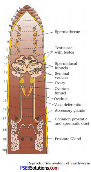
Question 3.
Draw a labelled diagram of the reproductive organs of an earthworm.
Answer:
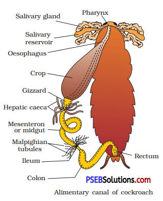
Question 4.
Draw a labelled diagram of alimentary canal of a cockroach.
Answer:
Question 5.
Distinguish between the following:
(a) Prostomium and peristomium.
(b) Septal nephridium and pharyngeal nephridium.
Answer:
(a) Prostomium and Peristomium: The first segment of earthworm with a ventral mouth is known as peristomium. Prostomium is a dorsal, lobe which is present on the ventral mouth.
(b) Septal Nephridia and Pharyngeal Nephridia: Septal nephridia are present on both the sides of intersegmental septa of segment 15 to the ‘ last that open into intestine.
The pharyngeal nephridia are closed (no nephrostome) nephridia present as three paired groups (of about 100) in 4th, 5th and 6th segments.
![]()
Question 6.
What are the cellular components of blood?
Answer:
The cellular components of blood are red blood cells, white blood cells and platelets.
Question 7.
What are the following and where do you find them in animal body (a) Chondrocytes (b) Axons (c) Ciliated epithelium.
Answer:
(a) Chondrocytes: These are the matrix secreting cells of the cartilage. These are found in the cartilage of connecting tissue.
(b) Axon: It is a long fibre, the distal end of which is branched. Each branch terminates as a bulb like structure called synaptic knob. The axon transmit nerve impulses away from the cell body.
(c) Ciliated Epithelium: If the columnar or cuboidal cells of columnar and cuboidal epithelium bear cilia on their free surface they are called ciliated epithelium.
Question 8.
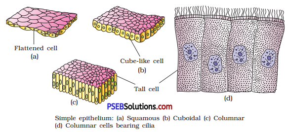
Describe various types of epithelial tissues with the help of labelled diagrams.
Answer:
Epithelial Tissues: Epithelial tissues provide covering to the inner and outer lining of various organs. There are following two types of epithelial tissues :
1. Simple epithelium: Simple epithelium is composed of a single layer of cells. It functions as a lining for body cavities, ducts and tubes.
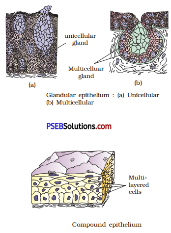
2. Compound epithelium: The compound epithelium consists of two or more cell layers. It has protective function as it does in our skin. It covers the dry surface of the skin, the moist surface of buccal cavity, pharynx, inner lining of ducts of salivary glands and of pancreatic ducts.
On the basis of structural modifications of the cells, simple epithelium is further divided into three types. These are :
(a) Squamous epithelium: The squamous epithelium is made of a single thin layer of flattened cells with irregular boundaries. They are found in the walls of blood vessels and air sacs of lungs and are involved in functions like forming a diffusion boundary.
(b) Cuboidal epithelium: The cuboidal epithelium is composed of a single layer of cube-like cells. This is commonly found in the ducts of glands and tubular parts of nephrons in kidneys. Its main functions are secretion and absorption.
(c) Columnar epithelium: The columnar epithelium is composed of a single layer of tall and slender cells. They are found in the lining of stomach and intestine and help in absorption and secretion.
(i) When the columnar or cuboidal cells bear cilia on their free surface they are called ciliated epithelium. Their function is to move particles or mucus in a specific direction over the epithelium organs like bronchioles and fallopian tubules.
(ii) Some of the columnar or cuboidal cells get specialized for secretion are called glandular epithelium. They are unicellular and multicellular.
![]()
Question 9.
Distinguish between:
(a) Simple epithelium and compound epithelium
(b) Cardiac muscle and striated muscle
(c) Dense regular and dense irregular connective tissue
(d) Adipose tissue and blood tissue
(e) Simple gland and compound gland
Answer:
(a) Differences between Simple and Compound Epithelium
| Simple Epithelium | Compound Epithelium |
| 1. It is composed of a single layer of | It consists of two or more cell layers. |
| 2. It functions as a lining for body cavities, ducts and tubes. | It is protective in function like our skin. |
(b) Differences between Cardiac and Striated Muscle
| Cardiac Muscle | Striated Muscle |
| 1. It occurs only in the wall of heart. | It occurs in the body wall, limb, tongue, pharynx, etc. |
| 2. They are short and cylindrical with truncate ends. | They are long and cylindrical with blunt ends. |
| 3. They have nerve supply from brain and autonomous nerve system | They have nerve supply from central nervous system. |
(c) Differences between Dense Regular and Dense Irregular Connective Tissues
| Dense Regular Connective Tissue | Dense Irregular Connective Tissue |
| Collagen fibres are present in rows between many parallel bundle of fibres. Examble: Tendons |
Fibroblasts and many fibre are present that are oriented differently. Examble: Cartilage, bones and blood. |
(d) Differences between Adipose Tissue and Blood Tissue
| Adipose Tissue | Blood Tissue |
| 1. It is a soft gel like connective tissue. | It is a fluid connective tissue. |
| 2. It is partitioned into lobules by septa. | There are no partitions. |
| 3. It is a storage tissue. | It is a transport tissue. |
| 4. Matrix is secreted by the cells. | Matrix is not secreted by the cells. |
| 5. It contain fibres. | Fibres are not conspicuous. |
| 6. Adipocytes contain fat droplets. | No cell of the tissue contains fat droplets. |
(e) Differences between Simple and Compound Gland
| Simple Gland | Compound Gland |
| 1. These glands have single unbranched duct. | These glands have branched system of ducts. |
| 2. These may be simple tubular glands, simple coiled tubular glands and simple alveolar glands. | These may be compound tubular glands, compound alveolar glands |
![]()
Question 10.
Mark the odd one in each series:
(a) Areolar tissue, blood, neuron, tendon
(b) RBC, WBC, platelets, cartilage
(c) Exocrine, endocrine, salivary gland; ligament
(d) Maxilla, mandible, labrum, antennae
(e) Protonema, mesothorax, metathorax, coxa
Answer:
(a) Neuron,
(b) Cartilage,
(c) Ligament,
(d) Antennae,
(e) Protonema.
Question 11.
Match the terms in column I with those in column II.
| Column I | Column II |
| A. Compound epithelium | 1. Alimentary canal |
| B. Compound eye | 2. Cockroach |
| C. Septal nephridia | 3. Skin |
| D. Open circulatory system | 4. Mosaic vision |
| E. Typhlosole | 5. Earthworm |
| F. Osteocytes | 6. Phallomere |
| G. Genitalia | 7. Bone |
Answer:
| Column I | Column II |
| A. Compound epithelium | 3. Skin |
| B. Compound eye | 4. Mosaic vision |
| C. Septal nephridia | 5. Earthworm |
| D. Open circulatory system | 2. Cockroach |
| E. Typhlosole | 1. Alimentary canal |
| F. Osteocytes | 7. Bone |
| G. Genitalia | 6. Phallomere |
Question 12.
Mention briefly about the circulatory system of earthworm.
Answer:
Pheretima exhibits a closed type of blood vascular system, consisting of blood vessels, capillaries and heart. Due to closed circulatory system, blood is confined to the heart and blood vessels. Contractions keep blood circulating in one direction. Smaller blood vessels supply the gut, nerve cord, and the body wall. Blood glands are present on the 4th, 5th and 6th segments. They produce blood cells and haemoglobin which is dissolved in blood plasma. Blood cells are phagocytic in nature. Earthworms lack specialised breathing devices. Respiratory exchange occurs through moist body surface into their blood stream.
![]()
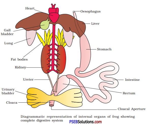
Question 13.
Draw a neat diagram of digestive system of frog.
Answer:
Question 14.
Mention the function of the following:
(a) Ureters in frog
(b) Malpighian tubules
(c) Body wall in earthworm
Answer:
(a) Functions of Ureters in Frog: In male frog, two ureters emerge from the kidneys. The ureters act as urinogenital duct which opens into the cloaca. Thus, the ureters carry both sperms and excretory wastes to the cloaca. In female frog, the ureters and oviduct open separately in the cloaca. The ureters in frog, thus acts as carrier of sperms and ova.
(b) Functions of Malpighian Tubules of Cockroach: Excretion is carried out by Malpighian tubules. Each tubule is lined by glandular cells. They absorb excretory waste products and converts them into uric acid which is excreted out through the hindgut.
(c) Functions of Body Wall of Earthworm: The body wall of earthworm has five layers – cuticle, epidermis, circular muscle layer, longitudinal muscle layer, peritoneum.
- Cuticle is a non-cellular elastic layer.
- The columnar cells of body wall provide support and therefore, are also known as supporting cells.
- Epidermis also has gland cells, receptor cells and basal cells.
- The glandular cell secrete mucus and thus, keep the skin moist, this
help in cutaneous respiration. - The last layer of the body wall is the outer membrane of the coelom called coelomic epithelium. The various muscle layers of the body wall provide strength and rigidity.