Punjab State Board PSEB 11th Class Biology Book Solutions Chapter 20 Locomotion and Movement Textbook Exercise Questions and Answers.
PSEB Solutions for Class 11 Biology Chapter 20 Locomotion and Movement
PSEB 11th Class Biology Guide Locomotion and Movement Textbook Questions and Answers
Question 1.
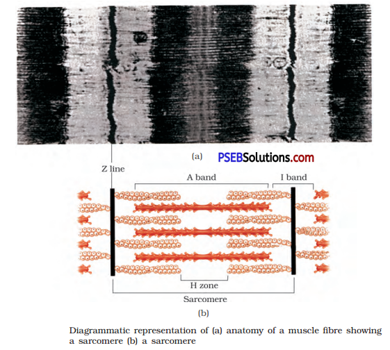
Draw the diagram of a sarcomere of skeletal muscle showing different regions.
Answer:
Question 2.
Define sliding-filament theory of muscle contraction.
Answer:
The sliding-filament, theory states that the contraction of a muscle fibre takes place by the sliding of the thin filaments over the thick filaments.
Question 3.
Describe the important steps in muscle contraction.
Answer:
Mechanism of Muscle Contraction:
- The mechanism of muscle contraction is explained by the sliding filament theory.
- This theory states that contraction of a muscle fibre is due to the sliding of the thin (actin) filaments over the thick (myosin) filaments.
- Muscle contraction is initiated by a neural signal from the central nervous system through a motor neuron.
- When the neural signal reaches the neuromuscular junction, it releases a neurotransmitter, i.e., acetylcholine, which generates an action potential in the sarcolemma.
- This spreads through the muscle fibre and causes the release of Ca++ ions from the sarcoplasmic reticulum into the sarcoplasm.
- The Ca++ ions bind to the subunit of troponin and brings about conformational changes; this removes the masking of the active site for myosin.
- The myosin head binds to the active site on actin to form a cross-bridge; this utilises energy from the hydrolysis of ATP.
- This pulls the actin filaments towards the centre of A-band.
- As a result, the Z-lines limiting the sarcomere are pulled closer together, causing a shortening of the sarcomere or contraction of muscle.
- Thus, during muscle contraction, the length of A band remains unchanged, while that of I-band decreases.
- The myosin goes back to its relaxed state.
- A new ATP binds and the cross-bridge is broken and the actin filaments slide out of A-band.
- The cycle of cross bridge-formation and cross bridge breakage continues till the Ca++ ions are pumped back to the sarcoplasmic reticulum which leads to the masking of the active site on F-actin.
![]()
Question 4.
Write true or false. If false change the statement so that it is true.
(a) Actin is present in thin filament.
(b) H-zone of striated muscle fibre represents both thick and thin filaments.
(c) Human skeleton has 206 bones.
(d) There are 11 pairs of ribs in man.
(e) Sternum is present on the ventral side of the body.
Answer:
(a) True
(b) False, H-zone represents thick filaments
(c) True
(d) False, there are 12 pairs of ribs in man.
(e) True
Question 5.
Write the difference between:
(a) Actin and Myosin
(b) Red and White Muscles
(c) Pectoral and Pelvic Girdle
Answer:
(a) Differences between Actin and Myosin Filament
| Actin Filaments | Myosin Filaments |
| 1. These are found in I-band. | These are found in A-band. |
| 2. These are thin. | These are thick. |
| 3. Cross bridges (heads) are absent. | Cross bridges (heads) are present. |
| 4. It is a globular protein with low molecular weight. | It is a heavy molecular weight polymerised protein. |
(b) Differences between Red and White Muscles
| Red Muscles | White Muscles |
| 1. In some muscles, myoglobin content is high, which gives a reddish colour to them, such muscles are called red muscles. | Some muscles possess very less quantity of myoglobin, so they appear whitish called as white muscles. |
| 2. These contain plenty of mitochondria. | These have less number of mitochondria but amount of sarcoplasmic reticulum is high. |
| 3. These are called aerobic muscles. | They depend on anaerobic process of energy. |
(c) Differences between Pectoral and Pelvic Girdle
| Pectoral Girdle | Pelvic Girdle |
| 1. It helps in the articulation of upper limbs. | It helps in the articulation of lower limbs. |
| 2. It is situated in the pectoral region of the body. | It is situated in the pelvic region of the body. |
| 3. Each half of pectoral girdle is formed of a clavicle and a scapula. | Pelvic girdle consists of two coxal bones. |
| 4. Scapula is a large triangular flat bone and clavicle is a long slender bone. | Each coxal bone is formed of three bones, ilium, ischium and pubis. |
| 5. An expanded process, acromion from scapula forms a depression called glenoid cavity, which articulates with the head of humerus to form shoulder joint. | Ilium, ischium and pubis fuse at a point to form a cavity called acetabulum to which the thigh bone articulates. |
Question 6.
Match Column-I with Column-II
| Column-I | Column-II |
| (a) Smooth muscle | (i) Myoglobin |
| (b) Tropomyosin | (ii) Thin filament |
| (c) Red muscle | (iii) Sutures |
| (d) Skull | (iv) Involuntary |
Answer:
| Column-I | Column-II |
| (a) Smooth muscle | (iv) Involuntary |
| (b) Tropomyosin | (ii) Thin filament |
| (c) Red muscle | (i) Myoglobin |
| (d) Skull | (iii) Sutures |
![]()
Question 7.
What are the different types of movements exhibited by the cells of human body?
Answer:
Cells of the human body exhibit three main types of movements-amoeboid, ciliary and muscular.
(i) Amoeboid Movement: Some specialised cells in our body like macrophages and leucocytes in blood exhibit amoeboid movement. It is effected by pseudopodia formed by the streaming of protoplasm (as in Amoeba). Cytoskeletal elements like microfilaments are also involved in amoeboid movement.
(ii) Ciliary Movement: Ciliary movement occurs in most of our internal tubular organs which are lined by ciliated epithelium. The coordinated movements of cilia in the trachea help us in removing dust particles and some of the foreign substances inhaled along with the atmospheric air. Passage of ova through the female reproductive tract is also facilitated by the ciliary movement.
(iii) Muscular Movement: Movement of our limbs, jaws, tongue, etc., require muscular movement. Locomotion requires a perfect coordinated activity of muscular, skeletal and neural systems.
Question 8.
How do you distinguish between a skeletal muscle and a cardiac muscle?
Answer:
| Skeletal Muscle | Cardiac Muscle |
| 1. The, cells of skeletal muscles are unbranched. | 1. The cells of cardiac muscles are branched. |
| 2. Intercalated disks are absent. | 2. The cells are joined with one another by intercalated disks that help in coordination or synchronization of the heartbeat. |
| 3. Alternate light and dark bands are present. | 3. Faint bands are present. |
| 4. They are voluntary muscles. | 4. They are involuntary muscles. |
| 5. They contract rapidly and get fatigued in a short span of time. | 5. They contract rapidly but do not get fatigued easily. |
| 6. They are present in body parts such as the legs, tongue, hands, etc. | 6. These muscles are present in the heart and control the contraction and relaxation of the heart. |
Question 9.
Name the type of joint between the following:
(i) Atlas/Axis
(ii) Carpal/Metacarpal of thumb
(iii) Between phalanges
(iv) Femur/Acetabulum
(v) Between cranial bones
(vi) Between pubic bones in the pelvic girdle.
Answer:
(i) Pivot joint
(ii) Saddle joint
(iii) Hinge joint
(iv) Ball and socket joint
(v) Fibrous joint
(vi) Cartilagenous joint
Question 10.
Fill in the blank spaces.
(a) All mammals (except a few) have ………………………………. cervical vertebra.
(b) The number of phalanges in each limb of human is ……………………………………
(c) Thin filament of myofibril contains 2 ‘F’ actins and two other proteins namely ………………………. and ………………..
(d) In a muscle fibre Ca2+ is stored in ………………………..
(e) ………………….. and ……………………………….. pairs of ribs are called floating ribs.
(f) The human cranium is made of …………………………. bones.
Answer:
(a) seven
(b) fourteen.
(c) troponin and tropomyosin
(d) sarcoplasm
(e) 11 th; 12th
(f) eight