Punjab State Board PSEB 11th Class Biology Book Solutions Chapter 21 Neural Control and Coordination Textbook Exercise Questions and Answers.
PSEB Solutions for Class 11 Biology Chapter 21 Neural Control and Coordination
PSEB 11th Class Biology Guide Neural Control and Coordination Textbook Questions and Answers
Question 1.
Briefly describe the structure of the following:
(a) Brain
(b) Eye
(c) Ear
Answer:
(a) Brain
The human brain has the following parts :
(i) Cerebrum
A deep cleft called longitudinal fissure divides the brain/cerebrum into two halves-cerebral hemispheres.
The two cerebral hemispheres are joined together by bundles of densely packed nerve fibers, called corpus callosum.
The outer surface of cerebrum, the cerebral cortex, is called grey matter, due to its greyish appearance; the cell bodies of the neurons are concentrated in this region; it contains motor areas, sensory areas and association areas.
Inner to the cortex is the white matter, that consists of myelinated nerve fibers in the form of nerve fibre tracts.
(ii) Thalamus
Thalamus is the major coordinating centre for sensory and motor signals.
(iii) Hypothalamus
It has centers to control body temperature, hunger, thirst, etc.
It contains several groups of neurosecretory cells, which secrete hormones.
(iv) Limbic System
The inner parts of the cerebral hemispheres and a group of deep structures called amygdala, hippocampus, etc. form a complex structure, called limbic system. Along with the hypothalamus, it is involved in the regulation of sexual behavior, expression of emotions, motivation, etc.
(v) Midbrain
Midbrain is located between the hypothalamus of the forebrain and the pons of the hindbrain. The dorsal portion of the midbrain consists of four small lobes, called corpora quadrigemina. A canal, called cerebral aqueduct passes through the midbrain.
(vi) Hindbrain
It consists of pons, cerebellum and medulla oblongata. The medulla contains centres which control vital functions like respiration, cardiovascular reflexes and gastric secretions. The medulla continues down as the spinal cord.
(b) Eye
- Each eye ball consists of three concentric layers, the outermost sclera, middle choroid and innermost retina.
- The sclera in the front (l/6th) forms the transparent cornea.
- The middle choroid is highly vascular and pigmented, that prevents internally reflected light within the eye; just behind the junction between cornea and sclera, the choroid becomes thicker forming the ciliary body.
- The iris extends from the ciliary body in front of the lens; it controls the dilation or constriction of pupil.
- The lens is suspended from the ciliary body, by suspensory ligaments.
- The anterior chamber of eye is filled with an aqueous clear fluid, aqueous humor and the posterior chamber has a gelatinous material, vitreous humor.
- The retina is composed of three layers of cells; the photoreceptor layer contains rods and cones, the intermediate layer has bipolar neurons, which synapse with retinal ganglion cells and their axons bundle to form optic nerve.
- The photoreceptor cells (rods and cones) contain the light sensitive proteins, called photopigments.
- The point in the retina where the optic nerve leaves the eye and the retinal blood vessels enter the eye is called a blind spot; there are no photoreceptor cells in this region.
- Lateral to blindspot, there is a yellowish pigmented spot, called macula lutea with a central pit called fovea.
- The fovea is the region where only cones are densely packed and it is the point where acuity (resolution) vision is the greatest.
(c) Ear
The ear performs two sensory functions, namely
(a) hearing and
(b) maintenance of body balance.
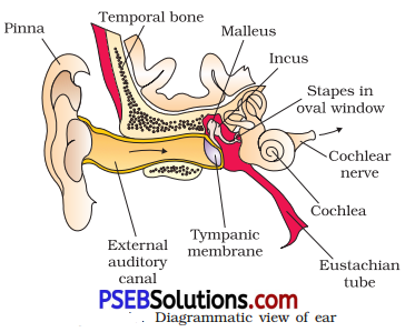
- Ear consists of three parts: external ear, middle ear and internal ear.
- The external ear consists of the pinna, and external auditory meatus.
- The tympanic membrane separates the middle ear from the external ear.
- The middle ear (tympanic cavity) is an air- filled chamber, which is connected to pharynx by Eustachian tube.
- The middle ear lodges three small bones, the ear ossicles namely, the malleus, incus and stapes.
- The middle ear communicates with the internal ear through the oval window and round window.
- The inner ear is a fluid-filled chamber and called labyrinth; it has two parts, an outer bony labyrinth, inside
- which a membranous labyrinth is floating in the perilymph; the membranous labyrinth is filled with a fluid, called endolymph.
- The labyrinth is divided into two parts, the cochlea and vestibular apparatus.
- Cochlea is the coiled portion of the labyrinth and its membranes, Reissner’s membrane and basilar membrane divide the perilymph-filled bony labyrinth into an upper scala vestibule, middle scala media and a lower scala tympani; scala media is filled with endolymph.
- At the base of the cochlea, scala vestibuli ends at the oval window, while the scala tympani terminates at the round window, that opens to the middle ear.
- Organ of Corti is the structural unit of hearing; it consists of hair cells which are the auditory receptors and is located on the basilar membrane.
- A thin elastic tectorial membrane lies over the row of hair cells.
- The vestibular apparatus is composed of three semicircular canals and an otolith organ or vestibule.
- The otolith organ has two parts namely the utricle and saccule.
- The utricle and saccule also contain a projecting ridge, called macula.
- The crista ampullar and macula are the specific receptors of the vestibular apparatus, for maintaining body balance.
![]()
Question 2.
Compare the following:
(a) Central Neural System (CNS) and Peripheral Neural System (PNS)
(b) Resting potential and action potential
(c) Choroid and retina
Answer:
(a) Comparison between Central Neural System (CNS) and Peripheral Neural System (PNS): The CNS includes the brain and the spinal cord and is the site of information processing and control. The PNS comprises of all the nerves of the body associated with the CNS (brain and spinal cord). The nerve fibers of the PNS are of two types :
(i) Afferent fibers, (ii) Efferent fibers
(b) Comparison between Resting Potential and Action Potential:
The electrical potential difference across the resting plasma membrane A is called the resting potential. The electrical potential difference across the plasma membrane at the site A is called the action potential, which is in fact termed as a nerve impulse.
(c) Comparison between Choroid and Retina: The middle layer of eyeball which contains many blood vessels and looks bluish in colour is known as choroid. The choroid layer is thin over the posterior two-thirds of the eyeball, but it becomes thick in the anterior part to form the ciliary body. The ciliary body itself continues forward to form a pigmented and opaque structure called the iris.
Retina is the inner layer of eye ball and it contains three layers of cells from inside to outside, i. e., ganglion cells, bipolar cells and photoreceptor cells. There are two types of photoreceptor cells, namely, rods and cones. These cells contain the light-sensitive proteins called the photopigments.
Question 3.
Explain the following processes:
(a) Polarisation of the membrane of a nerve fibre
(b) Depolarisation of the membrane of a nerve fibre
(c) Conduction of a nerve impulse along a nerve fibre
(d) Transmission \of a nerve impulse across a chemical synapse
Answer:
(a) Polarisation of the Membrane of a Nerve Fibre: During resting condition, the concentration of K+ ions is more inside the axoplasm while the concentration of Na+ ions is more outside the axoplasm. As a result, the potassium ions move faster from inside to outside as compared to sodium ions. Therefore, the membrane becomes positively charged outside and negatively charged inside. This is known as polarisation of membrane or polarised nerve.
(b) Depolarisation of the Membrane of a Nerve Fibre: When an electrical stimulus is given to a nerve fibre, an action potential is generated. The membrane becomes permeable to sodium ions than to potassium ions. This results into positive charge inside and negative charge outside the nerve fibre. Hence, the membrane is said to be depolarised.
(c) Conduction of a Nerve Impulse Along a Nerve Fibre: There are two types of nerve fibers-myelinated and non-myelinated. In myelinated nerve fibre, the action potential is conducted from node to node in jumping manner. This is because the myelinated nerve fibre is coated with myelin sheath.
The myelin sheath is impermeable to ions. As a result, the ionic exchange and depolarization of nerve fiber is not possible along the whole length of nerve fiber. It takes place only at some point, known as nodes of Ranvier, whereas in non-myelinated nerve fiber, the ionic exchange and depolarization of nerve fiber takes place along the whole length of the nerve fiber. Because of this ionic exchange, the depolarised area becomes repolarised and the next polarised area becomes depolarised.
(d) Transmission of a Nerve Impulse Across a Chemical Synapse:
Synapse is a small gap that occurs between the last portion of the axon of one neuron and the dendrite of next neuron. When an impulse reaches at the endplate of axon, vesicles consisting of chemical substances or neurotransmitters, such as acetylcholine, fuse with the plasma membrane.
This chemical moves across the cleft and attaches to chemo-receptors present on the membrane of the dendrite of next neuron. This binding of chemical with chemo-receptors leads to the depolarization of membrane and generates a nerve impulse across nerve fibre. The chemical, acetylcholine, is inactivated by enzyme acetylcholinesterase. The enzyme is present in the postsynaptic membrane of the dendrite. It hydrolyses acetylcholine and this allows the membrane to repolarise.
Question 4.
Draw labeled diagrams of the following:
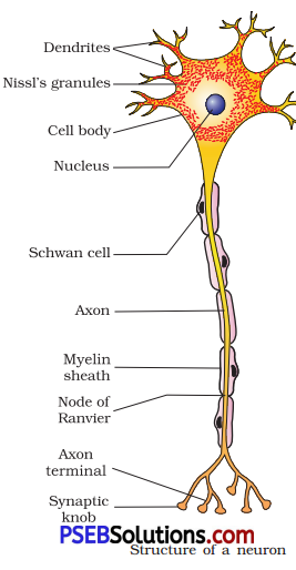
(a) Neuron
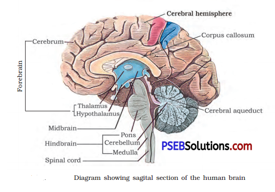
(b) Brain
(c) Eye
(d) Ear
Answer:
(a) Neuron
(b) Brain
(c) Eye

(d) Ear
Question 5.
Write short notes on the following:
(a) Neural coordination
(b) Forebrain
(c) Midbrain
(d) Hindbrain
(e) Retina
(f) Ear ossicles
(g) Cochlea
(h) Organ of Corti
(i) Synapse
Answer:
(a) Neural Coordination: The organized network of point-to-point connections for quick coordination provided by neural system is called neural coordination. The mechanism of neural coordination involves transmission of nerve impulses, impulse conduction across a synapse, and the physiology of reflex action.
(b) Forebrain: The forebrain consists of :
1. Olfactory lobes: The anterior part of the brain is formed by a pair of short club-shaped structures, the olfactory lobes. These are concerned with the sense of smell.
2. Cerebrum: It is the largest and most complex of all the parts of the human brain. A deep cleft divides the cerebrum longitudinally into two halves, which are termed as the left and right cerebral hemispheres connected by a large bundle of myelinated fibres the corpus callosum. The outer cover of cerebral hemisphere is called cerebral cortex. The cerebral cortex is referred to as the grey matter due to its greyish appearance (as neuron cell bodies are concentrated here).
The cerebral cortex is greatly folded. The upward folds, gyri, alternate with the downward grooves or sulci. Beneath the grey matter, there are millions of medullated nerve fibers, which constitute the inner part of the cerebral hemisphere. The large concentration of medullated nerve fibers gives this tissue an opaque white appearance. Hence, it is called the white matter.
3. Lobes: A very deep and a longitudinal fissure, separates the two cerebral hemispheres. Each cerebral hemisphere of the cerebrum is divided into four lobes, i.e., frontal, parietal, temporal, and occipital lobes.
In each cerebral hemisphere, there are three types of functional areas:
(i) Sensory areas receive impulses from the receptors and motor areas transmit impulses to the effectors.
(ii) Association areas are large regions that are neither clearly sensory nor motor injunction. They interpret the input, store the input and initiate a response in light of similar past experiences. Thus, these areas are responsible for complex functions like memory, learning, reasoning, and other intersensory associations.
(iii) Diencephalon is the posteroventral part of forebrain. Its main parts are as follows :
Epithalamus is a thin membrane of non-neural tissue. It is the posterior segment of the diencephalon. The cerebrum wraps around a structure called thalamus, which is a major coordinating center for sensory and motor signaling. The hypothalamus, that lies at the base of thalamus contains a number of centers, which control body temperature, urge for eating and drinking. It also contains several groups of neurosecretory cells, which secrete hormones called hypothalamic hormones.
(c) Midbrain: The midbrain is located between the thalamus and hypothalamus of the forebrain and pons of the hindbrain. A canal called the cerebral aqueduct passes through, the midbrain.
The dorsal portion of the midbrain mainly consists of two pairs (i.e., four) of rounded swellings (lobes) called corpora qua trigeminal.
(d) Hindbrain: The hindbrain consists of :
(i) Pons: It consists of fiber tracts that interconnect different regions of the brain.
(ii) Cerebellum: It is the second-largest part of the human brain (means little cerebrum). It has very convoluted surface in order to provide the additional space for many more neurons.
(iii) Medulla: It (oblongata) is connected to the spinal cord and contains centers, which control respiration, cardiovascular reflexes, and gastric secretions.
(e) Retina: The inner layer of eyeball is the retina and it contains three layers of cells from inside to outside—ganglion cells, bipolar cells and photoreceptor cells. There are two types of photoreceptor cells namely, rods and cones. These cells contain the light-sensitive proteins called the photopigments.
(f) Ear Ossicles: The middle ear contains three ossicles called malleus, incus and stapes which are attached to one another in a chain-like fashion. The malleus is attached to the tympanic membrane and the stapes is attached to the oval window or the cochlea. The ear ossicles increase the efficiency of transmission of sound waves to the inner ear.
(g) Cochlear: The membranous labyrinth of inner ear is filled with a fluid called endolymph. The coiled portion of the labyrinth is called cochlea. The membranes constituting cochlea, the Meissner’s and basilar, divide the surrounding perilymph-filled bony labyrinth into an upper scale vestibule and a lower scala tympani. The space within cochlea called scala media is filled with endolymph. At the base of the cochlea, the scala vestibule ends at the oval window, while the scala tympani terminates at the round window which opens to the middle ear.
(h) Organ of Corti: The organ of Corti is a structure located on the basilar membrane of inner ear, which contains hair cells that act as auditory receptors. The hair cells are present in rows on the internal side of the organ of Corti. The basal end of the hair cell is in close contact with the afferent nerve fibers. A large number of processes called stereocilia are projected from the apical part of each hair cell. Above the rows of the hair cells is a thin elastic membrane called tectorial membrane.
(i) Synapse: It is a junction between two neurons, where one neuron expands and comes in near contact with another neuron. A synapse is formed by the membranes of a pre-synaptic neuron, and a post-synaptic neuron, which may or may not be separated by a gap called synaptic cleft.
There are two types of synapses-an electrical synapse and a chemical synapse. In electrical synapse, membranes of pre and post-synaptic neurons are is very close proximity field. In chemical synapse, these membranes are separated by a fluid-filled space called synaptic cleft.
![]()
Question 6.
Give a brief account of:
(a) Mechanism of synaptic transmission
(b) Mechanism of vision
(c) Mechanism of hearing
Answer:
(a) Mechanism of Synaptic Transmission: Synapse is a junction between two neurons. It is present between the axon terminal of one neuron and the dendrite of next neuron separated by a cleft.
There are two ways of synaptic transmission :
1. Chemical transmission: When a nerve impulse reaches the end plate of axon, it releases a neurotransmitter (acetylcholine) across the synaptic cleft. This chemical is synthesized in cell body of the neuron and is transported to the axon terminal. The acetylcholine diffuses across the cleft and binds to the receptors present on the membrane of next neuron. This causes depolarization of membrane and initiates an action potential.
2. Electrical transmission: In this type of transmission, an electric current is formed in the neuron. This electric current generates an action potential and leads to transmission of nerve impulses across the nerve fiber. This represents a faster method of nerve conduction than the chemical method of transmission.
(b) Mechanism of Vision: Retina is the innermost layer of eye. It contains three layers of cells-inner ganglion cells, middle bipolar cells and outermost photoreceptor cells. A photoreceptor cell is composed of a protein called opsin and an aldehyde of vitamin A called retinal. When light rays are focused on the retina through cornea, it leads to the dissociation of retinal from opsin protein.
This changes the structure of opsin. As the structure of opsin changes, the permeability of membrane changes, generating a potential difference in the cells. This generates an action potential in the ganglionic cells and is transmitted to the visual cortex of the brain via optic nerves. In the cortex region of brain, the impulses are analyzed and image is formed on the retina.
(c) Mechanism of Hearing: The pinna of the external region collects the sound waves and directs it towards ear drum or external auditory canal. These waves strike the tympanic membrane and vibrations are created. Then, these vibrations are transmitted to the oval window, fenestra ovalis, through three ear ossicles, named as malleus, incus, and stapes. These ear ossicles act as lever and transmit the sound waves to internal ear.
These vibrations from fenestra ovalis are transmitted into cochlear fluid. This generates sound waves in the lymph. The formation of waves generates a ripple in the basilar membrane. This movement bends the sensory hair cells present on the organ of corti against tectorial membrane. As a result of this, sound waves are converted into nerve impulses. These impulses are then carried to auditory cortex of brain via auditory nerves. In cerebral cortex of brain, the impulses are analysed and sound is recognised.
Question 7.
Answer briefly:
(a) How do you perceive the colour of an object?
(b) Which part of our body helps us in maintaining the body balance?
(c) How does the eye regulate the amount of light that falls on the retina?
Answer:
(a) The daylight (photopic) vision and colour vision are functions of cones. In the human eye, there are three types of cones which possess their own characteristic photopigments that respond to red, green and blue lights. The sensations of different colours are produced by various combinations of these cones and their photopigments. When these cones are stimulated equally a sensation of white light is produced.
(b) The crista and macula are the specific receptors of the vestibular apparatus of inner ear which are responsible for the maintenance of balance of the body and posture.
(c) The diameter of the pupil is regulated by the muscle fiber of iris. Photoreceptors, rods, and cones regulate the amount of light that falls on the retina.
Question 8.
Explain the following:
(a) Role of Na+ in the generation of action potential.
(b) Mechanism of generation of light-induced impulse in the retina.
(c) Mechanism through which a sound produces a nerve impulse in the inner ear.
Answer:
(a) Role of Na+ in the Generation of Action Potential: When a stimulus is applied to a nerve, the membrane of the nerve becomes freely permeable to Na + . This leads to a rapid influx of Na+ followed by the reversal of the polarity at that site, i. e., the outer surface of the membrane becomes negatively charged and the inner side becomes positively charged. The electrical potential difference across plasma membrane at the membrane is called the action potential, which is in fact termed as a nerve impulse. Thus, this shows that Na+ ions play an important role in the conduction of nerve impulses.
(b) Mechanism of Generation of Light-induced Impulse in the Retina: Light induces dissociation of the retina from opsin resulting in changes in the structure of the opsin. This causes membrane permeability changes. As a result, potential differences are generated in the photoreceptor cells. This produces a signal that generates action potentials in the ganglion cells through the bipolar cells.
These action potentials (impulses) are transmitted by the optic nerves to the visual corted area of the brain, where the nerve impulses are analysed and the image formed on the retina is recognised.
(c) Mechanism through which a Sound Produces a Nerve Impulse in the Inner Ear: In the inner ear, the vibrations are passed through the oval window on to the fluid of the cochlea, where they generate waves in the lymph.
The waves in the lymphs induce a ripple in the basilar membrane.
These movements of the basilar membrane bend \ the hair cells, pressing them against the tectorial membrane. As a result, nerve impulses are generated in the associated afferent neurons. These impulses are transmitted by the afferent fibres via auditory nerves to the auditory cortex of the brain, where the impulses are analyzed and the sound is recognized.
Question 9.
Differentiate between:
(a) Myelinated and non-myelinated axons
(b) Dendrites and axons
(c) Rods and cones
(d) Thalamus and hypothalamus
(e) Cerebrum and cerebellum
Answer:
(a) Differences between Myelinated and Non-myelinated Axons
| Myelinated Axon | Non-myelinated Axon |
| 1. The myelinated nerve fibers are enveloped with Schwann cells, which form a myelin sheath around the axon. |
1. Unmyelinatcd nerve fibers are enclosed by a Schwann cell that does not form a myelin sheath around the axon. |
| 2. Myelinated nerve fibres are found in spinal and cranial nerves. | 2. They are commonly found in autonomous and the somatic neural systems. |
(b) Differences between Dendrite and Axon
| Dendrite | Axon |
| 1. These are short fibres which branch repeatedly and project out of the cell body also contain Nissl’s granules | The axon is a long branched fibre, which terminates as a bulb-like structure called. synaptic knob. It possess synaptic vesicles containing chemicals called neurotransmitters. |
| 2. These fibres transmit impulses towards the cell body. | The axons transmit nerve impulses away from the cell body to a synapse. |
(c) Differences between Rods and Cones
| Rod | Cone |
| 1. The twilight vision is the function of rods. | The daylight vision and colour vision are functions of cones. |
| 2. The rods contain a purplish-red protein called the rhodopsin or visual purple, which contains a derivative of Vitamin-A | In the human eye, there are three types of cones which possess their own characteristic photopigments that respond to red, green and blue lights. |
(d) Differences between Thalamus and Hypothalamus
| Thalamus | Hypothalamus |
| 1. The cerebrum wraps around a structure called thalamus. | It lies at the base of the thalamus. |
| 2. All types of sensory input passes synapses in the thalamus | It contains neurosecretory cells that secrete hypothalamus hormones. |
| 3. It controls emotional and memory functions. | It regulates, sexual behavior, expression of emotional reactions and motivation. |
.(e) Differences between Cerebrum and Cerebellum
| Cerebrum | Cerebellum |
| 1. It is the most developed part in brain. | It is the second developed part of brain also called as little cerebrum |
| 2. A deep cleft divides cerebrum into two cerebral hemispheres. | Externally the whole surface contains gyri and sulci. |
| 3. Its functions are intelligence, learning, memory, speech, etc. | It contains centres for coordination and error checking during motor and cognition. |
![]()
Question 10.
Answer the following:
(a) Which part of the ear determines the pitch of a sound?
(b) Which part of the human brain is the most developed?
(c) Which part of our central neural, system acts as a master clock?
Answer:
(a) Inner ear
(b) Cerebrum
(c) Brain
Question 11.
The region of the vertebrate eye, where the optic nerve passes out of the retina, is called the
(a) fovea
(b) iris
(c) blind spot
(d) optic chiasma
Answer:
(d) Optic Charisma
Question 12.
Distinguish between:
(a) Afferent neurons and efferent neurons.
(b) Impulse conduction in a myelinated nerve fibre and unmyelinated nerve fibre.
(c) Aqueous humour and vitreous humour.
(d) Blind spot and yellow spot.
(e) Cranial nerves and spinal nerves.
Answer:
(a) Differences between Afferent neurons and Efferent neurons
| Afferent Neurons | Efferent Neurons |
| The afferent nerve fibres transmit impulses from tissues/organs to the CNS. | The efferent fibres transmit regulatory impulses from the CNS to the concerned peripheral tissues/organs. |
(b) Differences between Myelinated and Non-myelinated Axons
| Myelinated Axon | Non-myelinated Axon |
| 1. The myelinated nerve fibers are enveloped with Schwann cells, which form a myelin sheath around the axon. |
1. Unmyelinatcd nerve fibers are enclosed by a Schwann cell that does not form a myelin sheath around the axon. |
| 2. Myelinated nerve fibres are found in spinal and cranial nerves. | 2. They are commonly found in autonomous and the somatic neural systems. |
(c) Differences between Aqueous humour and Vitreous humour
| Aqueous Humour | Vitreous Humour |
| 1. It is the space between the cornea and the lens. | The space between the lens and the retina is called the vitreous chamber. |
| 2. It contains a thin watery fluid. | It is filled with a transparent gel. |
(d) Differences between Blindspot and Yellow spot
| Blind Spot | Yellow Spot |
| 1. Photoreceptor cells are not present in this region. | Yellow spot or macula lutea is located at the posterior pole of the eye lateral to the blind spot. It has a central pit called fovea. |
| 2. The light focuses on that part of the retina is not detected. | The fovea of yellow spot is a thinned-out portion of retina where only the cones are densely packed is the point where visual cavity is greatest. |
(e) Differences between Cranial nerves and spinal nerves
| Cranial Nerves | Spinal Nerves |
| 1. The cranial nerves originate in the brain and terminate mostly in organs head and upper body. | The spinal nerves originate in the spinal cord and extend to parts of the body below the head. |
| 2. There are 12 pairs of cranial nerves. | There are 31 pairs of spinal nerves. |
| 3. Most of the cranial nerves contain axons and both sensory and motor neurons. | All of the spinal nerves contain axons of both sensory and motor neurons. |